140 posters, 10 videos, 15 audios, 20 sessions, 508 authors, 204 institutions
ePostersLive by SciGen Technologies S.A. All rights reserved.
8th World Congress on Vascular Access 2024
April 17 - 19, 2024 | Prague, Czech Republic

149
Unusual malposition of a peripherally inserted central catheter into the Anomalous left Brachiocephalic vein: A case report
Part of Topic
Unusual malposition of PICC into the Anomalous left Brachiocephalic vein: A case reportBackground
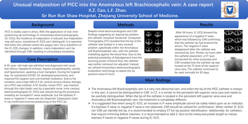
PICC is widely used in clinics. With the application of real-time positioning tip technology of intracavitary electrocardiography (IC-ECG), the incidence of malposition is reduced, but malposition may still occur, sometimes IC-ECG can't distinguish. It is reported that when the catheter enters the azygos vein, the p waveform of the IC-ECG changes. In addition, many malposition can't be identified through CXR, resulting in unnecessary extubation.
Case Description:
A 45-year-old male was admitted and diagnosed with acute liver failure, hepatitis B cirrhosis, hepatic encephalopathy, ascites, and hypertension, awaiting a liver transplant. During his hospital stay, he contracted COVID-19, developed pneumonia, and required ICU support and oral tracheal intubation. Due to his condition and a high INR of 3.16, central venous infusion therapy via a PICC was initiated. A 5F double-lumen PICC was inserted through the right basilic vein by a specialist nurse. Intra-cavitary electrocardiography (IC-ECG) was utilized during the procedure, revealing an increased P-wave amplitude, but the biphasic P-wave or negative P-wave was not observed. Subsequent chest X-ray confirmed incorrect positioning of the PICC tip.
Methods:
Despite initial electrocardiogram and CXR findings suggesting an atypical tip position, the catheter remained functional. Computed Tomography (CT) revealed that the tip of the catheter was located in a very unusual position, specifically within the Anomalous Left Brachiocephalic vein, with the catheter indwelling against the blood flow. Due to the potential possibility of dislodgement from the scouring power of blood flow, the catheter was neither removed nor adjusted. Instead, we utilized intracavitary electrocardiography localization technology to assess the tip position every 8 hours.
Results:
After 56 hours, IC-ECG showed the appearance of a negative P wave, which was followed by CXR confirming that the catheter tip had entered the atrium. The negative P wave disappeared after the catheter was retracted by 2cm. Ninety-six hours after the catheter placement, a CT scan (conducted for other purposes) and CXR revealed that the catheter tip was located at the T5 level of the superior vena cava. The catheter continued to be used normally for 92 days.
Main findings: