140 posters, 10 videos, 15 audios, 20 sessions, 508 authors, 204 institutions
ePostersLive by SciGen Technologies S.A. All rights reserved.
8th World Congress on Vascular Access 2024
April 17 - 19, 2024 | Prague, Czech Republic

141
In vitro and in vivo performance evaluation of a novel ultrasound-based vein visualization device
Poster Presenter
Authors
Michael Liddelow, Phuoc Hao Ho, Matthew Redknap, Cara Boyce, Katherine Arenson, Nicholas Buckley, Peter Carr, Barry Doyle, Nikhilesh Bappoo
Affiliations
VeinTech, School of Nursing and Midwifery, University of Galway, Vascular Engineering Laboratory, Harry Perkins Institute of Medical Research, QEII Medical Centre Nedlands and Centre for Medical Research, The University of Western Australia
Part of Topic
Video
In vitro and in vivo performance evaluation of a novel ultrasound-based vein visualization device
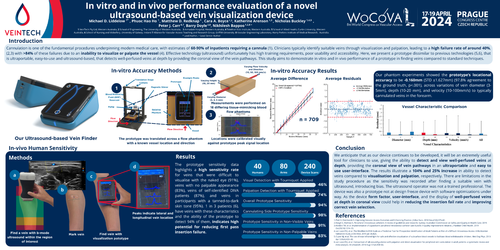
Cannulation is one of the fundamental procedures underpinning modern medical care, with estimates of 60-90%of inpatients requiring a cannula (1). Clinicians typically identify suitable veins through visualization and palpation, leading to a high failure rate of around 40%,(2,3) with >84% of these failures due to an inability to visualize or palpate the vessel (4). Effective technology (ultrasound) unfortunately has high training requirements, poor usability and accessibility. Here, we present a prototype dissimilar to previous technologies (5,6), that is ultraportable, easy-to-use and ultrasound-based, that detects well-perfused veins at depth by providing the coronal view of the vein pathways. This study aims to demonstrate in vitro and in vivo performance of a prototype in finding veins compared to standard techniques.
Our phantom experiments showed the prototype’s locational accuracy to be -0.160mm (STD ±1.627mm) (97.8% agreement to the ground truth, p<.001), across variations of vein diameter (3-5mm), depth (10-20 mm), and velocity (10-100mm/s) to typically cannulated veins in the forearm.
The prototype sensitivity data highlights a high sensitivity rate for veins that were difficult to visualise with the naked eye (91%), veins with no palpable appearance (83%), veins of self-identified DIVA patients (87%), and veins in participants with a tanned-to-dark skin tone (95%). 1 in 3 patients (6), have veins with these characteristics and the ability of the prototype to detect 94% of them, indicates high potential for reducing first pass insertion failure.
We anticipate that as our device continues to be developed, it will be an extremely useful tool for clinicians to use, giving the ability to detect and view well-perfused veins at depth, providing the coronal view of vein pathways in an ultraportable and easy to use user-interface. The results illustrate a 104% and 25% increase in ability to detect veins compared to visualisation and palpation, respectively. There are limitations in the study procedure as the sensitivity was recorded after finding a suitable vein with ultrasound, introducing bias. The ultrasound operator was not a trained professional. The device was also a prototype not at design freeze device with software optimizations under way. As the device form factor, user-interface, and the display of well-perfused veins at depth in coronal view could help in reducing the insertion fail rate and improving correct vein selection.