140 posters, 10 videos, 15 audios, 20 sessions, 508 authors, 204 institutions
ePostersLive by SciGen Technologies S.A. All rights reserved.
8th World Congress on Vascular Access 2024
April 17 - 19, 2024 | Prague, Czech Republic

129
Challenges of central venous access start with successful insertion
Poster Presenter
Part of Topic
CHALLENGES OFCENTRALVENOUS ACCESSSTART WITHSUCCESSFUL INSERTION
K.POLÁŠKOVÁ, M.KÝR, T.MERTA, O.ROHLEDER
Department of Pediatric Oncology, University Hospital Brno
Introduction:
Pediatric oncological patients have to undergo many procedures as intravenous treatment, imaging with intravenous contrast media, parenteral nutrition or repeated blood tests. Long term central venous access is often key of successful oncological treatment. On the other hand wide range of complications from mechanical, through hematological to infectious can occur and some of these complications can be life threatening for the patients
Case report:
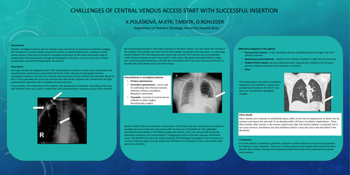
An 8-year-old boy was diagnosed with T-NHL lymphoblastic lymphoma. Initial tumor involvement was supraclavicular, paratracheal, and mediastinal lymph nodes. Because of anticipated intensive oncological treatment, insertion of a centrally inserted central venous catheter was indicated. Based on the CT scan supraclavicular access for insertion was safe and the catheter was inserted from the right supraclavicular approach with tunneling to the pectoral area.
In two months, the malfunction of the catheter with paravasation manifested. According to the X-ray with contrast, there was a leak in several sites and some depositions along the course of the catheter.
We assumed the damage or more likely presence of the fibrin sheath. This was reason for removal of the catheter. The catheter was intact but the fibrin sheath remained in the vasculature. In a few days second catheter from left supraclavicular access was inserted. The insertion was uncomplicated under ultrasound guidance and X-ray control. In two weeks, the patient developed febrile, cough, pain, and attenuated breathing on the left side. According to the X-ray, there was pneumothorax on the left side and fluidothorax to 1/3 of the thorax.
Based on MDCT there was extensive condensation of the lower lobe and marked volume reduction, probably because of extensive pneumonia with the presence of fluidothorax. The radiologist described discrete defects in the filling of segmental arteries, and it was not possible to exclude pulmonary embolism, but unfortunately CT angiography could not be done because of technical issues. The fluidothorax had to be solved surgically. Microbiological investigation did not prove any concrete infectious agent, but the patient was still febrile and therefore he was covered with wide spectrum antibiotics.
Differential diagnosis in this patient:
This complications was almost completely resolved on the antibiotics, surgical and symptomatic treatment. On the CT scan, there are only discrete radiological changes.
Conclusion:
For some patients, especially in pediatrics, long term central catheters are necessary equipment for intensive cancer treatment. There are no many evidence what happen with most of the fibrin sheaths after catheter removal, but healthcare professionals need to be prepared and evaluate many options.