141 posters, 8 videos, 8 audios, 5 topics, 442 authors, 228 institutions
ePostersLive by SciGen Technologies S.A. All rights reserved.
6th World Congress on Vascular Access 2021
April 29 - 30, 2021 | Virtual

P063
Tunneled permanent hemodialysis catheter placement in a patient with two cardiac implantable devices
Poster Presenter
Authors
Elisa Deganello, Elisa Deganello, Carlo Crepaldi, Fabio Baratto, Domenico Montemurro
Affiliations
Part of Topic
Research & Innovations
Tunneled permanent hemodialysis catheter placement in a patient with two cardiac implantable devices
A. Malagoli, E. Deganello, C. Crepaldi, F. Baratto, D. Montemurro.
Ospedali Riuniti Padova Sud, Monselice, Italy
Case description
The clinical case we describe concerns a 77-year-old patient with chronic renal failure, type 2 insulin dependent diabetes mellitus and sick sinus syndrome that required implantation of a permanent pacemaker.
The subject had been hospitalized for congestive heart failure and anuric renal failure and needed dialysis treatment through temporary CVC implantation in the right internal jugular vein.
For failure to resume renal function, the patient needed permanent vascular access to continue chronic hemodialysis treatment.
The heart disease (right ventricular dilatation, moderate mitral regurgitation and pulmonary hypertension), the late referral status on dialysis and the use of both subclavian veins for implantation of two pacemarkers in the last 20 years (first right, then left) justified the choice of tunneled permanent CVC for hemodialysis to replace the temporary CVC in the right internal jugular vein as the definitive vascular access for hemodialysis.
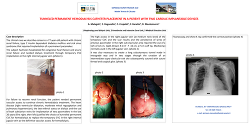
The high access in the right jugular vein (at medium neck level) of the temporary CVC and the cicatrizal results and the persitance of wires of previous pacemarker in the right subclavicle area required the use of a CVC of 32 cm, (Split-Stream R 14 F x 32 cm, 27 cm cuff tip, medCOMP) normally used in the left jugular vein. It was also necessary to create a long subcutaneous tunnel made in retrograde way and in two stages through the creation of an intermediate supraclavicle exit site subsequently sutured with suture thread and surgical glue. Fluoroscopy and chest X ray confirmed the correct position (photo 1)