875 posters, 25 topics, 3,440 authors, 1,061 institutions
ePostersLive by SciGen Technologies S.A. All rights reserved.
SAGES 2026 Annual Meeting Emerging Technology Session
March 25-28, 2026 | Tampa, FL, USA

P379
Cholecystitis in the Setting of Gallbladder Duplication
Poster Presenter
Authors
Affiliations
Part of Topic
Biliary
Cholecystitis in the Setting of Gallbladder Duplication
Cody Crnkovic, MD; Reagan M. Setser, MD; Irada Ibrahim-zada, MD
BACKGROUND
There are several reported variants in biliary anatomy. In literature, complete duplication of the gallbladder is exceedingly rare.1,2
Here, we report of a case of cholecystitis in a patient with complete gallbladder duplication with isolated involvement of one of the gallbladders
CASE PRESENTATION
A 27-year-old male with past medical history of nephrolithiasis presented to the Emergency Department for evaluation of abdominal pain for three days. His pain was localized to the right abdomen and worsened with meals. He had tenderness to palpation in his right upper quadrant. Lab workup revealed no leukocytosis or hyperbilirubinemia. A CT scan was obtained, which demonstrated complete gallbladder duplication with inflammation of the more anterior gallbladder, concerning for cholecystitis.
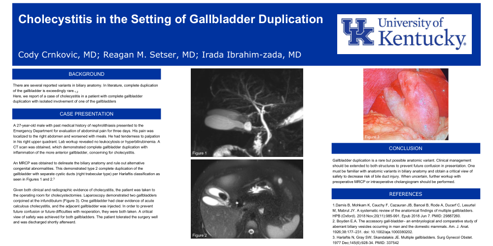
An MRCP was obtained to delineate the biliary anatomy and rule out alternative congenital abnormalities. This demonstrated type 2 complete duplication of the gallbladder with separate cystic ducts (right trabecular type) per Harlaftis classification as seen in Figures 1 and 2.3
Given both clinical and radiographic evidence of cholecystitis, the patient was taken to the operating room for cholecystectomies. Laparoscopy demonstrated two gallbladders conjoined at the infundibulum (Figure 3). One gallbladder had clear evidence of acute calculous cholecystitis, and the adjacent gallbladder was injected. In order to prevent future confusion or future difficulties with reoperation, they were both taken. A critical view of safety was achieved for both gallbladders. The patient tolerated the surgery well and was discharged shortly afterward.
CONCLUSION
Gallbladder duplication is a rare but possible anatomic variant. Clinical management should be extended to both structures to prevent future confusion in presentation. One must be familiar with anatomic variants in biliary anatomy and obtain a critical view of safety to decrease risk of bile duct injury. When uncertain, further workup with preoperative MRCP or intraoperative cholangiogram should be performed.
REFERENCES
1.Darnis B, Mohkam K, Cauchy F, Cazauran JB, Bancel B, Rode A, Ducerf C, Lesurtel M, Mabrut JY. A systematic review of the anatomical findings of multiple gallbladders. HPB (Oxford). 2018 Nov;20(11):985-991. Epub 2018 Jun 7. PMID: 29887260.
2. Boyden E.A. The accessory gall-bladder– an embryological and comparative study of aberrant biliary vesicles occurring in man and the domestic mammals. Am. J. Anat. 1926;38:177–231. doi: 10.1002/aja.1000380202.
3. Harlaftis N, Gray SW, Skandalakis JE. Multiple gallbladders. Surg Gynecol Obstet. 1977 Dec;145(6):928-34. PMID: 337542