875 posters, 25 topics, 3,440 authors, 1,061 institutions
ePostersLive by SciGen Technologies S.A. All rights reserved.
SAGES 2026 Annual Meeting Emerging Technology Session
March 25-28, 2026 | Tampa, FL, USA

P123
PRIMARY OMENTAL ABSCESS WITHOUT INTRA-ABDOMINAL SOURCE: A CASE REPORT AND SYSTEMATIC REVIEW
Poster Presenter
Authors
Affiliations
Part of Topic
Acute Care
Case Presentation:
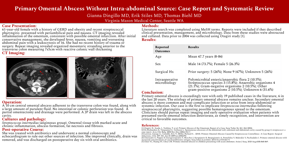
61-year-old female with a history of GERD and obesity and recent streptococcal pharyngitis presented with periumbilical pain and nausea. CT imaging revealed inflammation of the omentum, consistent with possible omental infarction. After initial conservative management, she developed fever, nausea, vomiting and worsening abdominal pain with a leukocytosis of 16. She had no recent history of trauma or surgery. Repeat imaging revealed organized mesenteric stranding anterior to the transverse colon measuring 7x5cm with reactive colonic wall thickening
CT Imaging:
Operation:
A 10 cm central omental abscess adherent to the transverse colon was found, along with a large amount of purulent fluid. No intestinal or colonic perforation was found. A partial omentectomy and drainage were performed. A JP drain was left in the abscess cavity.
Cultures and pathology:
Streptococcus intermedius (anginosus group). Omental tissue with marked acute and chronic inflammation, abscess formation, fat necrosis and fibrosis.
Post-operative Course:
She was treated with antibiotics and underwent a normal colonoscopy and echocardiogram to rule out other sources of infection. She improved clinically, drain was removed, and was discharged on postoperative day six with oral antibiotics.
Methods:
Literature search was completed using MeSH terms. Reports were included if they described clinical presentation, management, and microbiology. Data from these studies were abstracted and collated. Data prior to 2004 was collected using Otagiri study [1].
Results:
Conclusion:
Primary omental abscess is exceedingly rare with only 19 published cases in the literature, 6 in the last 20 years. The etiology of primary omental abscess remains unclear. Secondary omental abscess is more common and may complicate infarction or arise from intra-abdominal or systemic infection. Our case is the first to implicate Streptococcus intermedius following streptococcal pharyngitis, suggesting possible hematogenous spread to the omentum. Clinicians should pursue repeat imaging and early operative evaluation when patients with presumed sterile omental infarction deteriorate, as timely recognition, and intervention are critical to favorable outcomes.