2,281 posters, 63 videos, 56 audios, 43 topics, 18 sessions, 6,174 authors, 2,323 institutions
ePostersLive by SciGen Technologies S.A. All rights reserved.
RCOG World Congress 2025
June 23 - 25, 2025 | ExCeL London, UK
EP.0863
Atypical Placental site nodule, a case series report:
Poster Presenter
Authors
Affiliations
Part of Topic
Gynaecological oncology
Atypical Placental site nodule, a case report:
Objective:
We would like to raise awareness of this condition, which presents like any other common pathology so that early diagnosis and prompt treatment can resolve symptoms.
Case Series reports:
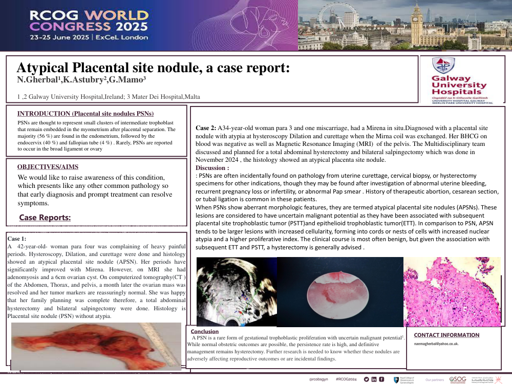
Case 1 : A 42-year-old- woman para four was complaining of heavy painful periods. Hysteroscopy, Dilation, and curettage were done and histology showed an atypical placental site nodule (APSN). Her periods have significantly improved with Mirena. However, on MRI she had adenomyosis and a 6cm ovarian cyst. On computerized tomography(CT ) of the Abdomen, Thorax, and pelvis, a month later the ovarian mass was resolved and her tumor markers are reassuringly normal. She was happy that her family planning was complete therefore, a total abdominal hysterectomy and bilateral salpingectomy were done. Histology is Placental site nodule (PSN) without atypia.
Case 2: A34-year-old woman para 3 and one miscarriage, had a Mirena in situ.Diagnosed with a placental site nodule with atypia at hysteroscopy Dilation and curettage when the Mirna coil was exchanged. Her BHCG on blood was negative as well as Magnetic Resonance Imaging (MRI) of the pelvis. The Multidisciplinary team discussed and planned for a total abdominal hysterectomy and bilateral salpingectomy which was done in November 2024 , the histology showed an atypical placenta site nodule
Discussion: PSNs are thought to represent small clusters of intermediate trophoblast that remain embedded in the myometrium after placental separation. The majority (56 %) are found in the endometrium, followed by the endocervix (40 %) and fallopian tube (4 %) . Rarely, PSNs are reported to occur in the broad ligament or ovary . These are often incidentally found on pathology from uterine curettage, cervical biopsy, or hysterectomy specimens for other indications, though they may be found after investigation of abnormal uterine bleeding, recurrent pregnancy loss or infertility, or abnormal Pap smear . History of therapeutic abortion, cesarean section, or tubal ligation is common in these patients.
When PSNs show aberrant morphologic features, they are termed atypical placental site nodules (APSNs). These lesions are considered to have uncertain malignant potential as they have been associated with subsequent placental site trophoblastic tumor (PSTT)and epithelioid trophoblastic tumor(ETT). In comparison to PSN, APSN tends to be larger lesions with increased cellularity, forming into cords or nests of cells with increased nuclear atypia and a higher proliferative index. The clinical course is most often benign, but given the association with subsequent ETT and PSTT, a hysterectomy is generally advised .
Conclusion:
A PSN is a rare form of gestational trophoblastic proliferation with uncertain malignant potential1. While normal obstetric outcomes are possible, the persistence rate is high, and definitive management remains hysterectomy. Further research is needed to know whether these nodules are adversely affecting reproductive outcomes or are incidental findings.
Objective:
We would like to raise awareness of this condition, which presents like any other common pathology so that early diagnosis and prompt treatment can resolve symptoms.