2,281 posters, 63 videos, 56 audios, 43 topics, 18 sessions, 6,174 authors, 2,323 institutions
ePostersLive by SciGen Technologies S.A. All rights reserved.
RCOG World Congress 2025
June 23 - 25, 2025 | ExCeL London, UK
EP.0855
Synchronous Endometrioid Adenocarcinomas Ovary and Endometrium in Young Woman: A Case Report
Poster Presenter
Authors
Affiliations
Part of Topic
Gynaecological oncology
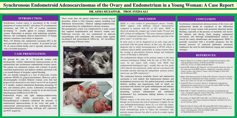
Synchronous Endometroid Adenocarcinomas of the Ovary and Endometrium in a Young Woman: A Case Report
INTRODUCTION:
Synchronous ovarian cancer is uncommon in the overall population of women with EC, with about 5% of endometrial carcinomas and 10% to 20% of ovarian carcinomas developing.1 It usually appear in younger, nulliparous women. Particularly in patients with underlying metabolic illnesses, the overlapping clinical characteristics of benign ailments sometimes cause delays in diagnosis.
With a 4% lifetime risk, endometrial carcinoma (EC) is the most prevalent pelvic cancer in women. It accounts for just 2% of cancer-related deaths and is typically detected early with a favorable prognosis.2
CASE PRESENTATION:
We present the case of a 35-year-old woman with histologically verified endometrioid adenocarcinoma of the endometrium and synchronous endometrioid ovarian cancer. The patient was unmarried woman with a history of poorly controlled hypertension and diabetes mellitus presented with a six-month history of irregular vaginal bleeding.
She was initially managed as a case of polycystic ovarian syndrome (PCOS) by general practitioners. However, pelvic ultrasonography revealed bilateral ovarian cysts that had progressively increased in size from 4 cm to 8.8 cm in period of 6 months, marked endometrial thickening measuring 25 mm, and minimal pelvic ascites. Laboratory investigations showed normal tumor markers, except for an elevated lactate dehydrogenase (LDH) level of 302 U/L.
Endometrial biopsy and right salpingo-oophorectomy performed with fertility preservation in mind. Histopathological evaluation confirmed the presence of endometrioid adenocarcinoma of the ovary and grade 1 endometrioid adenocarcinoma of the endometrium, with immunohistochemistry revealing wild-type p53 expression and WT1 negativity.
Three weeks later, the patient underwent a second surgical procedure, which is total clearance surgery including total abdominal hysterectomy, bilateral salpingo-oophorectomy, and omentectomy for comprehensive disease clearance.
Her postoperative course was complicated by a septic wound that required hospitalization and intensive wound care. Following recovery, she was commenced on adjuvant chemotherapy with carboplatin. She remains under regular oncological and gynecological follow-up for surveillance and monitoring of disease status.
DISCUSSION
SEOC is a rare variant of gynecological cancers. Usually, SEOC appears with different clinical characteristics compared to patients with EC or OC alone. SEOC is observed among the younger age women under 55 years and 40% of them are nulliparous.3 The most common symptom of SEOC is abnormal uterine bleeding and pelvic pain due to the pelvic mass3,4 as with this case.
In most cases it can be diagnosed at an early stage and is associated with low-grade disease. In this case, the delay in diagnosis due to initial mismanagement as PCOS reflects a common clinical pitfall, particularly in young women where the overlap in presentation between benign and malignant causes of bleeding is substantial.
The endometrioid subtype of the primary tumors is the most common histological finding with the rate of 50%-70% of cases. In our report, both women with SEOC had endometrioid histological type2 , usually they are low grade (1&2) that do not metastasize earlier and are associated with favorable outcome showing the independent primary tumors and in our case H/P confirmed it.5
The connection between metabolic factors and endometrial carcinoma is well studied, that hypertension diabetes and dyslipidemia increases risk. Our patient had poorly controlled diabetes mellitus and hypertension both of which are associated with chronic low-grade inflammation and immune dysfunction, impairing innate immune responses and promoting systemic inflammation and endothelial dysfunction.6 key mechanism linking diabetes to tumor aggressiveness.
Primary surgery is the main stray of treatment7 but adjuvant therapy given because the risk of metastasis is higher. In our case immunohistochemistry shows P53 +ve wild type tumor, which is WT –ve. Owing to aggressive nature, carboplatin based chemotherapy given to reduce the recurrence. Our patients remains disease free after 6 cycles & is under regular surveillance
CONCLUSION
Synchronous endometrioid adenocarcinoma of the ovary and endometrium should be considered in the differential diagnosis of young women with persistent abnormal uterine bleeding, especially in the presence of metabolic risk factors like diabetes and obesity. Early imaging, endometrial assessment, and thorough histopathological analysis are crucial for timely identification and management. SEOC is generally associated with a favorable prognosis, but the unusual presence of suspected pulmonary metastasis emphasizes the need for individualized staging and treatment strategies.
REFERENCES: