2,281 posters, 63 videos, 56 audios, 43 topics, 18 sessions, 6,174 authors, 2,323 institutions
ePostersLive by SciGen Technologies S.A. All rights reserved.
RCOG World Congress 2025
June 23 - 25, 2025 | ExCeL London, UK
EP.0853
Incidental Finding of Epithelioid Trophoblastic Tumor During Fertility Evaluation: A Case Report
Poster Presenter
Part of Topic
Gynaecological oncology
Incidental Finding of Epithelioid Trophoblastic Tumor During Fertility Evaluation: A Case Report
Case Description
Authers: Razan Abukhaizaran1, Mohand Abulihya2, Omar Abeddayem1, Mira Abu Samra1,3, Mashhour Naasan4
Affiliations: (1) Razan Center for Infertility, Palestine , (2) Istishari Arab Hospital, Ramallah, Palestine, (3) Specialized Arab Hospital, Nablus, Palestine, (4) Arab Hospitals Group, Palestine
Objective
To report a rare case of epithelioid trophoblastic tumor (ETT) incidentally diagnosed during fertility evaluation, emphasizing its clinical presentation, diagnostic challenges, and management.
A 36-year-old Patient (G1P1A0) presented to Razan Center for Infertility in October 2024 for frozen embryo transfer using embryos from a previous IVF/ICSI cycle.
Fertility History: Unexplained infertility since 2010; one IVF/ICSI cycle in 2014 (5 frozen day-2 embryos); spontaneous pregnancy in 2023 ended in preterm delivery and neonatal death in March 2023.
Current complaint: Irregular vaginal bleeding.
Investigations:
TVUS: Thickened endometrium
β-hCG: Negative
Hysteroscopy: Suspicious lesion on the posterior uterine wall
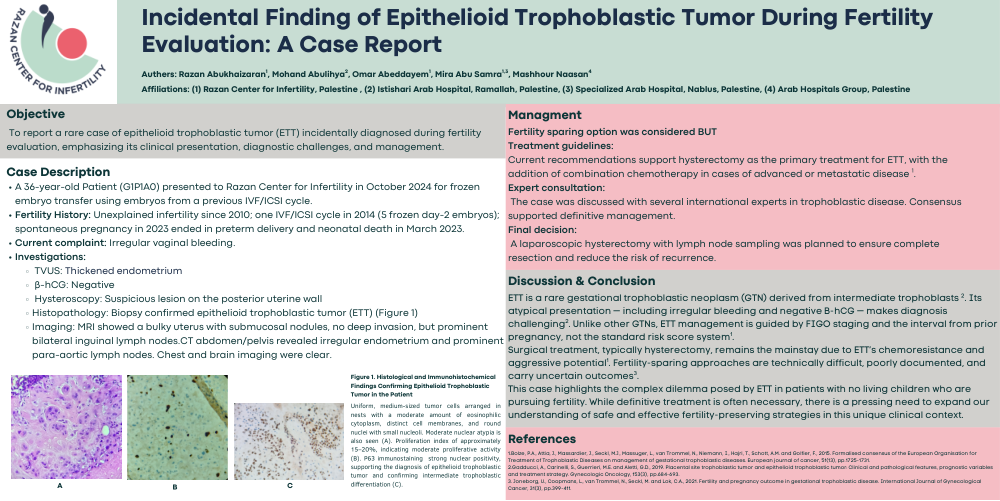
Histopathology: Biopsy confirmed epithelioid trophoblastic tumor (ETT) (Figure 1)
Imaging: MRI showed a bulky uterus with submucosal nodules, no deep invasion, but prominent bilateral inguinal lymph nodes.CT abdomen/pelvis revealed irregular endometrium and prominent para-aortic lymph nodes. Chest and brain imaging were clear.
Figure 1. Histological and Immunohistochemical Findings Confirming Epithelioid Trophoblastic Tumor in the Patient
Uniform, medium-sized tumor cells arranged in nests with a moderate amount of eosinophilic cytoplasm, distinct cell membranes, and round nuclei with small nucleoli. Moderate nuclear atypia is also seen (A). Proliferation index of approximately 15–20%, indicating moderate proliferative activity (B). P63 immunostaining strong nuclear positivity, supporting the diagnosis of epithelioid trophoblastic tumor and confirming intermediate trophoblastic differentiation (C).
Managment
Fertility sparing option was considered BUT
Treatment guidelines:
Current recommendations support hysterectomy as the primary treatment for ETT, with the addition of combination chemotherapy in cases of advanced or metastatic disease 1.
Expert consultation:
The case was discussed with several international experts in trophoblastic disease. Consensus supported definitive management.
Final decision:
A laparoscopic hysterectomy with lymph node sampling was planned to ensure complete resection and reduce the risk of recurrence.
Discussion & Conclusion
ETT is a rare gestational trophoblastic neoplasm (GTN) derived from intermediate trophoblasts 2. Its atypical presentation — including irregular bleeding and negative β-hCG — makes diagnosis challenging2. Unlike other GTNs, ETT management is guided by FIGO staging and the interval from prior pregnancy, not the standard risk score system1.
Surgical treatment, typically hysterectomy, remains the mainstay due to ETT’s chemoresistance and aggressive potential1. Fertility-sparing approaches are technically difficult, poorly documented, and carry uncertain outcomes3.
This case highlights the complex dilemma posed by ETT in patients with no living children who are pursuing fertility. While definitive treatment is often necessary, there is a pressing need to expand our understanding of safe and effective fertility-preserving strategies in this unique clinical context.