2,281 posters, 63 videos, 56 audios, 43 topics, 18 sessions, 6,174 authors, 2,323 institutions
ePostersLive by SciGen Technologies S.A. All rights reserved.
RCOG World Congress 2025
June 23 - 25, 2025 | ExCeL London, UK
EP.0844
DIAGNOSTIC CHALLENGE IN A CASE OF TORTED HUGE OVARIAN FIBROMA WITH MEIG-SYNDROME
Poster Presenter
Authors
Affiliations
Part of Topic
Gynaecological oncology
Diagnostic Challenge In a Case of Torted Huge Ovarian Fibroma with Meig Syndrome
Case presentation
A woman in her 50s presented to the Emergency Department with a three-day history of severe, sharp, diffuse abdominal pain and multiple episodes of vomiting. She denied dyspnoea, fever, lower urinary tract symptoms or bowel abnormalities. The patient’s past medical history included depression.
On examination, the patient appeared significantly pale. Abdominal examination revealed distention, diffuse tenderness, and a non-reducible, tender, erythematous 7x6 cm periumbilical mass with overlying skin bruising. The patient was tachycardic, tachypnoeic, and hypotensive upon admission, with a normal body temperature and an oxygen saturation of 97% on room air.
Initial blood tests showed a haemoglobin of 66 g/L (not previously investigated or symptomatic), prompting packed red blood cell transfusions. White cell count was 10.8 x 10⁹/L, platelets were 187 x 109/L, and C-reactive protein was elevated at 198 mg/L. CA-125 was elevated at 413 U/mL. Electrolytes, liver function tests, and renal function tests were within normal range.
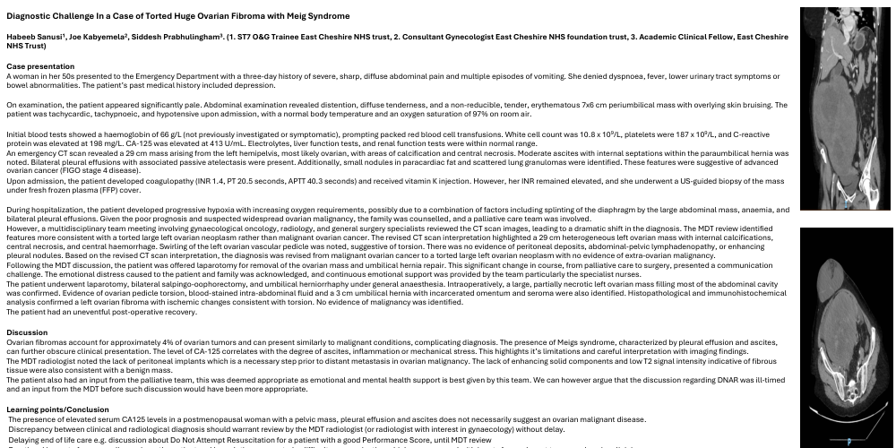
An emergency CT scan revealed a 29 cm mass arising from the left hemipelvis, most likely ovarian, with areas of calcification and central necrosis. Moderate ascites with internal septations within the paraumbilical hernia was noted. Bilateral pleural effusions with associated passive atelectasis were present. Additionally, small nodules in paracardiac fat and scattered lung granulomas were identified. These features were suggestive of advanced ovarian cancer (FIGO stage 4 disease).
Upon admission, the patient developed coagulopathy (INR 1.4, PT 20.5 seconds, APTT 40.3 seconds) and received vitamin K injection. However, her INR remained elevated, and she underwent a US-guided biopsy of the mass under fresh frozen plasma (FFP) cover.
During hospitalization, the patient developed progressive hypoxia with increasing oxygen requirements, possibly due to a combination of factors including splinting of the diaphragm by the large abdominal mass, anaemia, and bilateral pleural effusions. Given the poor prognosis and suspected widespread ovarian malignancy, the family was counselled, and a palliative care team was involved.
However, a multidisciplinary team meeting involving gynaecological oncology, radiology, and general surgery specialists reviewed the CT scan images, leading to a dramatic shift in the diagnosis. The MDT review identified features more consistent with a torted large left ovarian neoplasm rather than malignant ovarian cancer. The revised CT scan interpretation highlighted a 29 cm heterogeneous left ovarian mass with internal calcifications, central necrosis, and central haemorrhage. Swirling of the left ovarian vascular pedicle was noted, suggestive of torsion. There was no evidence of peritoneal deposits, abdominal-pelvic lymphadenopathy, or enhancing pleural nodules. Based on the revised CT scan interpretation, the diagnosis was revised from malignant ovarian cancer to a torted large left ovarian neoplasm with no evidence of extra-ovarian malignancy.
Following the MDT discussion, the patient was offered laparotomy for removal of the ovarian mass and umbilical hernia repair. This significant change in course, from palliative care to surgery, presented a communication challenge. The emotional distress caused to the patient and family was acknowledged, and continuous emotional support was provided by the team particularly the specialist nurses.
The patient underwent laparotomy, bilateral salpingo-oophorectomy, and umbilical herniorrhaphy under general anaesthesia. Intraoperatively, a large, partially necrotic left ovarian mass filling most of the abdominal cavity was confirmed. Evidence of ovarian pedicle torsion, blood-stained intra-abdominal fluid and a 3 cm umbilical hernia with incarcerated omentum and seroma were also identified. Histopathological and immunohistochemical analysis confirmed a left ovarian fibroma with ischemic changes consistent with torsion. No evidence of malignancy was identified.
The patient had an uneventful post-operative recovery.
Discussion
Ovarian fibromas account for approximately 4% of ovarian tumors and can present similarly to malignant conditions, complicating diagnosis. The presence of Meigs syndrome, characterized by pleural effusion and ascites, can further obscure clinical presentation. The level of CA-125 correlates with the degree of ascites, inflammation or mechanical stress. This highlights it’s limitations and careful interpretation with imaging findings.
The MDT radiologist noted the lack of peritoneal implants which is a necessary step prior to distant metastasis in ovarian malignancy. The lack of enhancing solid components and low T2 signal intensity indicative of fibrous tissue were also consistent with a benign mass.
The patient also had an input from the palliative team, this was deemed appropriate as emotional and mental health support is best given by this team. We can however argue that the discussion regarding DNAR was ill-timed and an input from the MDT before such discussion would have been more appropriate.
Learning points/Conclusion
- The presence of elevated serum CA125 levels in a postmenopausal woman with a pelvic mass, pleural effusion and ascites does not necessarily suggest an ovarian malignant disease.
- Discrepancy between clinical and radiological diagnosis should warrant review by the MDT radiologist (or radiologist with interest in gynaecology) without delay.
- Delaying end of life care e.g. discussion about Do Not Attempt Resuscitation for a patient with a good Performance Score, until MDT review
- Emotional impact of a cancer diagnosis on the patient and her relatives presented a difficult communication which was managed with inputs from relevant teams and senior clinicians