2,281 posters, 63 videos, 56 audios, 43 topics, 18 sessions, 6,174 authors, 2,323 institutions
ePostersLive by SciGen Technologies S.A. All rights reserved.
RCOG World Congress 2025
June 23 - 25, 2025 | ExCeL London, UK
EP.0841
A Case Report of Mesonephric Adenocarcinoma of Vagina in a Postmenopausal Lady
Poster Presenter
Part of Topic
Gynaecological oncology
Audio
Title: A Case Report of Mesonephric Adenocarcinoma of Vagina in a Postmenopausal Lady
E. Nnamani1, S. Boctor1, A. Cheung2, K. Taylor1
1.Department of Obstetrics and Gynaecology, University Hospital of Wales, Cardiff.
2. Department of Clinical Oncology, Velindre University NHS Trust, Wales.
INTRODUCTION:
Mesonephric adenocarcinoma of the vagina is an exceptionally rare tumor believed to arise from mesonephric (Wolffian) remnants. The diagnosis is often challenging due to its atypical immunohistochemical profile and nonspecific presentation. We present a unique case of a postmenopausal woman diagnosed with mesonephric adenocarcinoma, initially presenting as a ruptured vaginal cyst.
CASE REPORT:
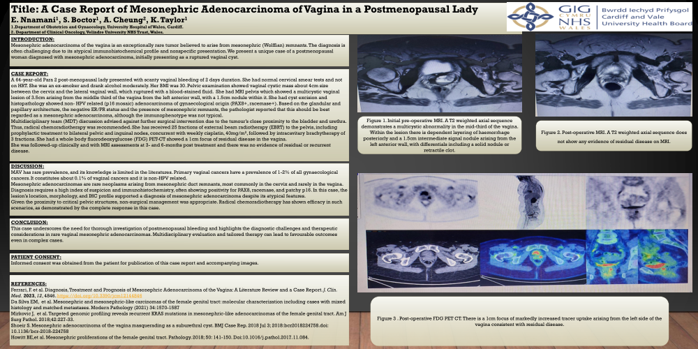
A 64-year-old Para 2 post-menopausal lady presented with scanty vaginal bleeding of 2 days duration. She had normal cervical smear tests and not on HRT. She was an ex-smoker and drank alcohol moderately. Her BMI was 30. Pelvic examination showed vaginal cystic mass about 4cm size between the cervix and the lateral vaginal wall, which ruptured with a blood-stained fluid. She had MRI pelvis which showed a multicystic vaginal lesion of 3.5cm arising from the middle third of the vagina from the left anterior wall, with a 1.5cm nodule within it. She had cyst excision and histopathology showed non- HPV related (p16 mosaic) adenocarcinoma of gynaecological origin (PAX8+, racemase+). Based on the glandular and papillary architecture, the negative ER/PR status and the presence of mesonephric remnants, the pathologist reported that this should be best regarded as a mesonephric adenocarcinoma, although the immunophenotype was not typical.
Multidisciplinary team (MDT) discussion advised against further surgical intervention due to the tumour’s close proximity to the bladder and urethra. Thus, radical chemoradiotherapy was recommended. She has received 25 fractions of external beam radiotherapy (EBRT) to the pelvis, including prophylactic treatment to bilateral pelvic and inguinal nodes, concurrent with weekly cisplatin, 40mg/m2, followed by intracavitary brachytherapy of 3 fractions. She had a whole body fluorodeoxyglucose (FDG) PET-CT showed a 1cm focus of residual disease in the vagina.
She was followed-up clinically and with MRI assessments at 3- and 6-months post treatment and there was no evidence of residual or recurrent disease.
DISCUSSION:
MAV has rare prevalence, and its knowledge is limited in the literatures. Primary vaginal cancers have a prevalence of 1-2% of all gynaecological cancers. It constitutes about 0.1% of vaginal cancers and it is non-HPV related.
Mesonephric adenocarcinomas are rare neoplasms arising from mesonephric duct remnants, most commonly in the cervix and rarely in the vagina. Diagnosis requires a high index of suspicion and immunohistochemistry, often showing positivity for PAX8, racemase, and patchy p16. In this case, the lesion’s location, morphology, and IHC profile supported a diagnosis of mesonephric adenocarcinoma despite its atypical features.
Given the proximity to critical pelvic structures, non-surgical management was appropriate. Radical chemoradiotherapy has shown efficacy in such scenarios, as demonstrated by the complete response in this case.
CONCLUSION:
This case underscores the need for thorough investigation of postmenopausal bleeding and highlights the diagnostic challenges and therapeutic considerations in rare vaginal mesonephric adenocarcinomas. Multidisciplinary evaluation and tailored therapy can lead to favourable outcomes even in complex cases.
PATIENT CONSENT:
Informed consent was obtained from the patient for publication of this case report and accompanying images.
REFERENCES:
Ferrari, F. et al. Diagnosis, Treatment and Prognosis of Mesonephric Adenocarcinoma of the Vagina: A Literature Review and a Case Report. J. Clin. Med. 2023, 12, 4846. https://doi.org/10.3390/jcm12144846
Da Silva EM, et al. Mesonephric and mesonephric-like carcinomas of the female genital tract: molecular characterization including cases with mixed histology and matched metastases. Modern Pathology (2021) 34:1570-1587
Mirkovic J, et al. Targeted genomic profiling reveals recurrent KRAS mutations in mesonephric-like adenocarcinomas of the female genital tract. Am J Surg Pathol. 2018;42:227-33.
Shoeir S. Mesonephric adenocarcinoma of the vagina masquerading as a suburethral cyst. BMJ Case Rep. 2018 Jul 3; 2018:bcr2018224758.doi: 10.1136/bcr-2018-224758
Howitt BE,et al. Mesonephric proliferations of the female genital tract. Pathology. 2018; 50: 141-150. Doi:10.1016/j.pathol.2017.11.084.
Figure 1. Initial pre-operative MRI. A T2 weighted axial sequence demonstrates a multicystic abnormality in the mid-third of the vagina. Within the lesion there is dependent layering of haemorrhage posteriorly and a 1.5cm intermediate signal nodule arising from the left anterior wall, with differentials including a solid nodule or retractile clot.
Figure 2. Post-operative MRI. A T2 weighted axial sequence does not show any evidence of residual disease on MRI.
Figure 3 . Post-operative FDG PET CT. There is a 1cm focus of markedly increased tracer uptake arising from the left side of the vagina consistent with residual disease.