2,281 posters, 63 videos, 56 audios, 43 topics, 18 sessions, 6,174 authors, 2,323 institutions
ePostersLive by SciGen Technologies S.A. All rights reserved.
RCOG World Congress 2025
June 23 - 25, 2025 | ExCeL London, UK
EP.0837
Primary extranodal marginal zone B-cell lymphoma in a macroscopically normal fallopian tube
Poster Presenter
Authors
Affiliations
Part of Topic
Gynaecological oncology
Primary extranodal marginal zone B-cell lymphoma in a macroscopically normal right fallopian tube
V Su1, J Wu1
1 Department of Obstetrics and Gynaecology, Royal Prince Alfred Hospital, Sydney, Australia
Objective:
To explore the role of chronic infection and/or autoimmune dysregulation in the lymphomagenesis of gynaecological extranodal marginal zone lymphoma (EMZL)
Case Report:
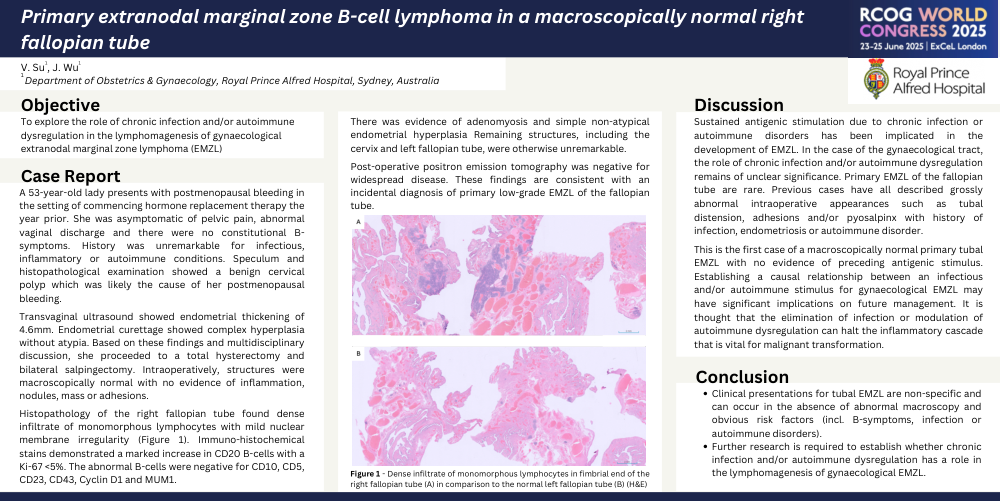
A 53-year-old lady presents with postmenopausal bleeding in the setting of commencing hormone replacement therapy the year prior. She was asymptomatic of pelvic pain, abnormal vaginal discharge and there were no constitutional B-symptoms. History was unremarkable for infectious, inflammatory or autoimmune conditions. Speculum and histopathological examination showed a benign cervical polyp which was likely the cause of her postmenopausal bleeding. Transvaginal ultrasound showed endometrial thickening of 4.6mm. Endometrial curettage showed complex hyperplasia without atypia. Based on these findings and multidisciplinary discussion, she proceeded to a total hysterectomy and bilateral salpingectomy. Intraoperatively, structures were macroscopically normal with no evidence of inflammation, nodules, mass or adhesions. Histopathology of the right fallopian tube found dense infiltrate of monomorphous lymphocytes with mild nuclear membrane irregularity (Figure 1). Immuno-histochemical stains demonstrated a marked increase in CD20 B-cells with a Ki-67 <5%. The abnormal B-cells were negative for CD10, CD5, CD23, CD43, Cyclin D1 and MUM1. There was evidence of adenomyosis and simple non-atypical endometrial hyperplasia, but remaining structures, including the cervix and left fallopian tube, were otherwise unremarkable. Post-operative positron emission tomography was negative for widespread disease. These findings are consistent with an incidental diagnosis of primary low-grade EMZL of the fallopian tube.
Discussion:
Sustained antigenic stimulation due to chronic infection or autoimmune disorders has been implicated in the development of EMZL. In the case of the gynaecological tract, the role of chronic infection and/or autoimmune dysregulation remains of unclear significance. Primary EMZL of the fallopian tube are rare. Previous cases have all described grossly abnormal intraoperative appearances such as tubal distension, adhesions and/or pyosalpinx with history of infection, endometriosis or autoimmune disorder. This is the first case of a macroscopically normal primary tubal EMZL with no evidence of preceding antigenic stimulus. Establishing a causal relationship between an infectious and/or autoimmune stimulus for gynaecological EMZL may have significant implications on future management. It is thought that the elimination of infection or modulation of autoimmune dysregulation can halt the inflammatory cascade that is vital for malignant transformation.
Conclusion:
Clinical presentations for tubal EMZL are non-specific and can occur in the absence of abnormal macroscopy and obvious risk factors (incl. B-symptoms, infection or autoimmune disorders). Further research is required to establish whether chronic infection and/or autoimmune dysregulation has a role in the lymphomagenesis of gynaecological EMZL.