2,281 posters, 63 videos, 56 audios, 43 topics, 18 sessions, 6,174 authors, 2,323 institutions
ePostersLive by SciGen Technologies S.A. All rights reserved.
RCOG World Congress 2025
June 23 - 25, 2025 | ExCeL London, UK
EP.0828
A Rare Case of Pyoderma Gangrenosum in a Postoperative Gynaecologic Oncology Patient
Poster Presenter
Authors
Rafeef Abu Shamleh, Ahmad Souriti, Patrick Keating, Nagindra Das
Affiliations
Part of Topic
Gynaecological oncology
Pyoderma Gangrenosum of the Abdominal Wall Skin in a Postoperative Gynaecologic Oncology Patient
R.Abu Shamleh, A.Souriti, P.Keating, N.Das, K. Lutchman-Singh (Members of Swansea Bay University Health Board, Gynaecology Oncology Department, United Kingdom)
Case presentation:
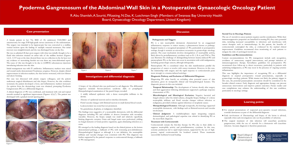
A female patient in her 70s, BMI of 40, underwent TAH/BSO and omentectomy for stage IVB high-grade serous carcinoma of the endometrium. The surgery was intended to be laparoscopic but was converted to a midline vertical incision upon the finding of multiple omental metastases. Her initial recovery was straightforward, and she was discharged home on day five.
She was re-admitted 28 days post-surgery with what was initially thought to be a florid skin infection situated on either side of the midline incision (Figures: 1,2,3). A CT scan at the time showed there was no collection in the fat layer nor any evidence of necrotising fasciitis nor was there any intra-abdominal sepsis. The cause of this was thought to be due to (LMWH) subcutaneous injections introducing infection into the skin.
Initial management was with IV antibiotics. Inflammatory markers were raised and wound swabs demonstrated Pseudomonas aeruginosa. However, despite clinical improvements in infection markers, the skin lesions worsened, with new blisters and ulcers forming.
Her case was discussed with plastic surgery colleagues, and the patient underwent surgical debridement and skin biopsy. However, the skin condition worsened, the patient developed a fever, and inflammatory markers increased. An opinion from the dermatology team was obtained, prompting Pyoderma Gangrenosum (PG) as a differential diagnosis.
A clinical diagnosis of PG was confirmed, and treatment with oral and topical steroids resulted in significant improvement (Figures: 4,5,6,7). The patient was discharged with a gradual steroid tapering plan.
Investigations and differential diagnosis:
A biopsy of the affected skin was performed to aid in diagnosis. The differential diagnoses included Stevens-Johnson syndrome (SJS) or pemphigoid. Histopathological examination of the punch biopsy revealed:
The findings were suggestive of a neutrophilic dermatosis, with the differential diagnoses including PG and cellulitis or abscess formation with secondary vasculitis. However, the biopsy sample was small and relatively superficial, limiting diagnostic certainty. Gram and fungal stains were performed, yielding no fungal elements or Gram-positive organisms, again eliminating infection as a cause.
PG emerged as the leading diagnosis based on the clinical picture as the lesions demonstrated pathergy, a hallmark of PG, with worsening post-debridement. Histopathological Support as although it is not definitive, the neutrophilic infiltrate and vascular changes were consistent with PG. This diagnosis was further supported by the patient’s response to corticosteroid therapy, a hallmark of PG management.
Discussion:
Pathogenesis and Triggers
PG is a rare neutrophilic dermatosis characterized by an exaggerated inflammatory response to minor trauma, a phenomenon known as pathergy. Surgical trauma is a recognized precipitant of PG, particularly in postoperative settings, though it remains an uncommon occurrence in gynaecologic oncology patients. The exact mechanism involves dysregulation of neutrophilic activity, likely driven by proinflammatory cytokines such as IL-1, IL-6, and TNF-α. This case illustrates the challenge of distinguishing between trauma-induced PG and paraneoplastic PG, as the latter can occur in association with solid malignancies, including gynaecologic cancers, although infrequently.
Paraneoplastic PG is considered when the skin manifestations parallel the progression of cancer or resolve with cancer treatment. However, in this case, the relationship with surgery and the rapid response to corticosteroids point more strongly to a trauma-induced aetiology.
Diagnostic Pathway and Exclusion of Differential Diagnoses
Diagnosing PG relies heavily on excluding other potential causes of skin ulceration, given its nonspecific clinical and histopathological features. The diagnostic process in this case included:
Treatment with Corticosteroids
Corticosteroids remain the first-line therapy for PG; due to their ability to suppress neutrophilic activity and proinflammatory cytokines. In this case, systemic prednisone led to rapid improvement, supported by the use of high-potency topical corticosteroids for localized control. These treatments successfully facilitated wound healing.
Steroid Use in Oncology Patients
The use of steroids in cancer patients requires careful consideration. While their immunosuppressive properties are beneficial in treating PG, they carry potential risks, including increased susceptibility to infections and interference with cancer therapies such as immunotherapy. In this patient, the benefits of corticosteroids outweighed the risks, as evidenced by the marked clinical improvement. Guidelines recommend close monitoring of such patients to mitigate the risks of prolonged steroid use.
Guidelines and Recommendations
Current recommendations for PG management include early recognition, avoidance of unnecessary surgical interventions, and prompt initiation of immunosuppressive therapy. Surveillance guidelines for postoperative PG recommend close follow-up to detect recurrence, particularly in patients with underlying malignancies, as PG may recur in the presence of ongoing systemic inflammation.
This case highlights the importance of recognizing PG as a differential diagnosis in atypical postoperative wound presentations, especially in gynaecologic oncology patients. While trauma was likely the primary trigger in this case, the potential paraneoplastic association should be considered in the broader clinical context. Early diagnosis and appropriate therapy can prevent unnecessary interventions and improve patient outcomes. Future studies and case compilations may enhance the understanding of this rare condition, particularly in oncology settings.
Learning points: