2,281 posters, 63 videos, 56 audios, 43 topics, 18 sessions, 6,174 authors, 2,323 institutions
ePostersLive by SciGen Technologies S.A. All rights reserved.
RCOG World Congress 2025
June 23 - 25, 2025 | ExCeL London, UK
EP.0824
METASTATIC PRIMARY OVARIAN PEComa: A CASE REPORT FROM DEDICATED CANCER HOSPITAL
Poster Presenter
Part of Topic
Gynaecological oncology
METASTATIC PRIMARY OVARIAN PEComa: A CASE REPORT
Iqra Yasin1, Afshan Saeed Usmani1, Aamir Ali Syed2
1 Fellow Gynaecologic Oncology, 2 Consultant Surgical Oncologist
PERIVASCULAR EPITHELIOID CELL TUMOR (PEComa)
• Rare mesenchymal neoplasm, characterized by expression of both melanocytic and smooth muscle markers. [1]
• Potential precursor lesion: minute perivascular epithelioid cell nests occurring in lymph nodes. [2]
• Associated with tuberous sclerosis complex (TSC) and frequently harbour TSC1/2 mutations including genetic pathway Rheb/mTOR/p70S6K (responsive to mTOR inhibitors). [3,4]
• Usually benign but some cases are malignant. [5]
• Common origin: abdominopelvic viscera especially GI and uterus. [6]
• Gynaecologic PEComas: uterine corpus, cervix, vagina, broad ligament, vulva and adnexa. [7]
• Ovarian PEComas: metastases of PEComa to the ovary, PEComatosis involving the ovary, [7] and primary ovarian PEComas (3 cases in literature). [8-10]
CASE SUMMARY
A 31-year-old female, married for 4 years, para 1 with 2 miscarriages and no comorbidities underwent excision of left adnexal mass outside SKMCH. Biopsy showed Perivascular epithelioid cell tumour (PEComa).
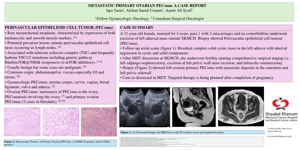
• Follow-up serial scans (figure 1): Residual complex solid cystic mass in the left adnexa with interval regression in cystic and solid components.
• After MDT discussion at SKMCH, she underwent fertility-sparing comprehensive surgical staging i.e., left salpingo-oophorectomy, excision of left pelvic wall mass excision, and infracolic omentectomy.
• Biopsy (Figure 2) showed left ovarian primary PEComa with metastatic deposits in the omentum and left pelvic sidewall.
• Case re-discussed in MDT: Targeted therapy is being planned after completion of pregnancy.
REFERENCES
1. Fletcher CD, Unni KK, Mertens F. Pathology and genetics of tumours of soft tissue and bone. Lyon: Iarc, 2002; 221-222.
2. Nagasaka T, Murakami Y, Sasaki E, Hosoda W, Nakanishi T, Yatabe Y. Minute perivascular epithelioid cell (PEC) nests in the abdominal lymph nodes—a putative precursor of PEComa. Pathol. Int. 2015; 65:193-6.
3. Ardeleanu C, Bussolati G. Telocytes are the common cell of origin of both PEComas and GISTs: an evidence‐supported hypothesis. J. Cell. Mol. Med. 2011; 15: 2569-74.
4. Martignoni G, Pea M, Reghellin D, Zamboni G, Bonetti F. PEComas: the past, the present and the future. Virchows Arch. 2008; 452: 119-32.
5. Folpe AL, Kwiatkowski DJ. Perivascular epithelioid cell neoplasms: pathology and pathogenesis. Hum. Pathol. 2010; 41: 1-5.
6. Doyle LA, Hornick JL, Fletcher CD. PEComa of the gastrointestinal tract: clinicopathologic study of 35 cases with evaluation of prognostic parameters. Am. J. Surg. Pathol. 2013; 37: 1769-82.
7. Conlon N, Soslow RA, Murali R. Perivascular epithelioid tumours (PEComas) of the gynaecological tract. J Clin Pathol. 2015; 68: 418-26.
8. Lee SE, Choi YL, Cho J, Kim T, Song SY, Sung CO. Ovarian perivascular epithelioid cell tumor not otherwise specified with transcription factor E3 gene rearrangement: a case report and review of the literature. Hum. Pathol. 2012; 43: 1126-30.
9. Anderson AE, Yang X, Young RH. Epithelioid angiomyolipoma of the ovary: a case report and literature review. Int. J. Gynecol. Pathol. 2002; 21: 69-73.
10. Westaby JD, Magdy N, Fisher C, El-Bahrawy M. Primary Ovarian Malignant PEComa: A Case Report. Int J Gynecol Pathol. 2017 Jul;36(4):400-404.