2,281 posters, 63 videos, 56 audios, 43 topics, 18 sessions, 6,174 authors, 2,323 institutions
ePostersLive by SciGen Technologies S.A. All rights reserved.
RCOG World Congress 2025
June 23 - 25, 2025 | ExCeL London, UK
EP.0720
30cm, 7kg cervical fibroid – A rare presentation of a common gynaecological problem
Poster Presenter
Authors
Samantha Maden, Sheriff Sheidu, Hala Selim, Edmund Inetianbor
Affiliations
Part of Topic
Gynaecological imaging
30cm, 7kg fibroid – A case report of a rare presentation of a common gynecological problem
Samantha Maden 1, Sheriff Sheidu1, Hala Selim2, Edmund Inetianbor2
1Specialist Trainee in Obstetrics and Gynaecology 2Consultant Obstetrician and Gynaecologist
Queen Elizabeth, the Queen Mother Hospital, Margate Kent
INTRODUCTION
Uterine fibroids, also known as leiomyomas, are common benign tumours in women of reproductive age, though they rarely reach the size and weight described in this case. Large fibroids during pregnancy can pose significant risks, including malposition and obstructed labour. We present an interesting case where an extremely large cervical fibroid was unexpectedly discovered at an elective myomectomy which was eventually managed via hysterectomy.
CASE REPORT
A 33-year-old nulliparous woman presented for her dating scan at 13 weeks of gestation. Ultrasound revealed an 18x13x16cm fibroid which was determined to be in the fundus of the uterus. Due to the large size of the fibroid, serial growth scans were sanctioned which showed growth of the fibroid with pregnancy. Due to the difficulty with obtaining adequate views, it was impossible to determine placental location until a transvaginal ultrasound was done at 36 weeks confirmed a posterior high placenta. Her final growth scan estimated that the fibroid was 19x17x16mm and that the baby was in a transverse lie.
At 39+ 5 weeks of gestation, she underwent a planned midline laparotomy for caesarean delivery due to the large size of fibroid and malpresentation. The baby was delivered in good condition. There were no complications and she made a routine postnatal recovery.
Without further imaging, she had open myomectomy to manage the fibroid. During this operation the known 19cm fibroid was removed from the fundus but unexpectedly, a larger cervical fibroid was discovered which occupied the whole of the lower pelvis. An attempt at removal of this fibroid could not be completed safely.
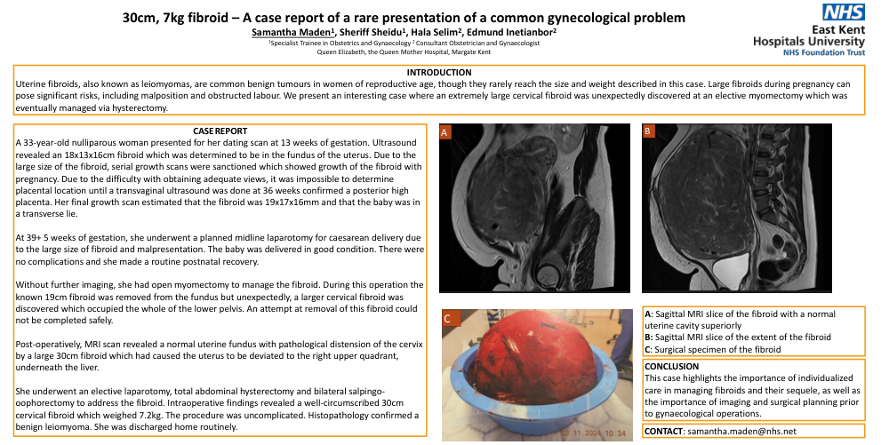
Post-operatively, MRI scan revealed a normal uterine fundus with pathological distension of the cervix by a large 28cm fibroid which had caused the uterus to be deviated to the right upper quadrant, underneath the liver.
She underwent an elective laparotomy, total abdominal hysterectomy and bilateral salpingo-oophorectomy to address the fibroid. Intraoperative findings revealed a well-circumscribed 30cm cervical fibroid which weighed 7.2kg. The procedure was uncomplicated. Histopathology confirmed a benign leiomyoma. She was discharged home routinely.
A: Sagittal MRI slice of the fibroid with a normal uterine cavity superiorly
B: Sagittal MRI slice of the extent of the fibroid
C: Surgical specimen of the fibroid
CONCLUSION
This case highlights the importance of individualized care in managing fibroids and their sequele, as well as the importance of imaging and surgical planning prior to gynaecological operations.