2,281 posters, 63 videos, 56 audios, 43 topics, 18 sessions, 6,174 authors, 2,323 institutions
ePostersLive by SciGen Technologies S.A. All rights reserved.
RCOG World Congress 2025
June 23 - 25, 2025 | ExCeL London, UK
EP.0622
Isolated anterior vaginal wall leomyoma in a postmenopausal woman
Poster Presenter
Authors
Affiliations
Part of Topic
General gynaecology
Video
CASE REPORT
Title: Isolated anterior vaginal wall leomyoma in a postmenopausal woman
Authors: Dr. Sunday Asogwa, Clinical fellow , Mr. Mohammed Elwerdany , OG Consultant
Department- Obstetrics & Gynaecology, James Paget University Hospital, Great Yarmouth
Objective: We present this rare diagnosis because of risk of misdiagnosis due to a number of commoner differentials.
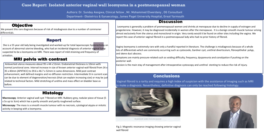
Report: She is a 55 year old lady being investigated and worked-up for total laparoscopic hysterectomy on account of abnormal uterine bleeding, who had an incidental diagnosis of anterior vaginal lump suspected to be a leomyoma on MRI. There was report of mild straining and frequency of urination.
MRI pelvis with contrast: Anteverted uterus measures about 95 x 44 x 51mm. Endometrial thickness is 10mm with normal junctional zone. Interval increase in size of known anterior vaginal wall fibroid from 26 x 35 x 40mm (APXTXCC) to 39.6 x 46.7 x 52mm in same dimensions. Mild post contrast enhancement, well defined margins and no diffusion restriction. Intermediate SI in current scan can be due to element of degeneration/necrosis (that can explain increasing size) or may be just related to technical factors. Mild stretching of urethra and mass effect on bladder base as before.
Histology:
Macroscopy: Anterior vaginal wall cyst ? fibroid on MRI. Rubbery grey, nodular piece of tissue (5 x 5x up to 3cm) which has a partly smooth and partly roughened surface.
Microscopy: The mass is a smooth muscle tumour with no necrosis, cytological atypia or mitotic activity in keeping with a leomyoma.
Discussion: Leomyoma is generally a problem of premenopausal women and shrinks at menopause due to decline in supply of estrogen and progesterone. However, it may be diagnosed incidentally in women after the menopause. It is a benign smooth muscle tumour arising almost exclusively from the uterus and monoclonal in origin. Very rarely would it be found on other sites including the vagina. We report this case of anterior vaginal fibroid in a postmenopausal lady who had no prior history of fibroid.
Vagina leomyoma is extremely rare with only a handful reported in literature. The challenge is misdiagnosis because of a whole lots of differentials which are commonly occurring such as cystocoele, Gardner cyst, urethral diverticulum, fibroepithelial polyp, and skene duct abscess.
Symptoms are mainly pressure-related such as voiding difficulty, frequency, dyspareunia and constipation if pushing on the posterior wall.
Excision is the main stay of management after intraoperative cystoscopy and urethral stenting to reduce the risk of injury.
Conclusions: Vaginal fibroid is a rarity and requires a high index of suspicion with the assistance of imaging such as MRI to make a diagnosis. Nevertheless, definitive diagnosis can only be reached following histology.