2,281 posters, 63 videos, 56 audios, 43 topics, 18 sessions, 6,174 authors, 2,323 institutions
ePostersLive by SciGen Technologies S.A. All rights reserved.
RCOG World Congress 2025
June 23 - 25, 2025 | ExCeL London, UK
EP.0620
Idiopathic Ovarian Vein Thrombosis: Early Recognition to Prevent Unnecessary Surgical Intervention
Poster Presenter
Authors
Affiliations
Part of Topic
General gynaecology
40-year-old multiparous woman presented with a 6-day history of severe left lower abdominal pain, nausea and fever. The pain was sharp, sudden in onset, continuous, and worsened by sitting.
She denied genitourinary symptoms, recent travel, prolonged immobility, oral contraception use or personal/family history of thrombosis.
Vital signs were stable.
She had localised left iliac fossa tenderness with rebound tenderness.
Bloods were unremarkable apart from raised C-reactive protein (236 mg/L)
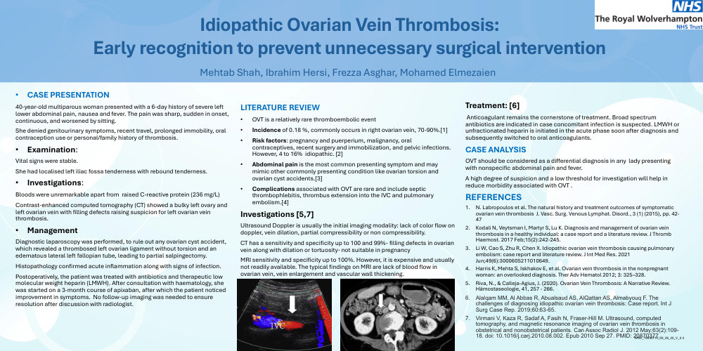
Contrast-enhanced computed tomography (CT) showed a bulky left ovary and left ovarian vein with filling defects raising suspicion for left ovarian vein thrombosis.
Diagnostic laparoscopy was performed, to rule out any ovarian cyst accident, which revealed a thrombosed left ovarian ligament without torsion and an edematous lateral left fallopian tube, leading to partial salpingectomy.
Histopathology confirmed acute inflammation along with signs of infection.
Postoperatively, the patient was treated with antibiotics and therapeutic low molecular weight heparin (LMWH). After consultation with haematology, she was started on a 3-month course of apixaban, after which the patient noticed improvement in symptoms. No follow-up imaging was needed to ensure resolution after discussion with radiologist.
LITERATURE REVIEW
Investigations [5,7]
Ultrasound Doppler is usually the initial imaging modality: lack of color flow on doppler, vein dilation, partial compressibility or non compressibility.
CT has a sensitivity and specificity up to 100 and 99%- filling defects in ovarian vein along with dilation or tortuosity- not suitable in pregnancy
MRI sensitivity and specificity up to 100%. However, it is expensive and usually not readily available. The typical findings on MRI are lack of blood flow in ovarian vein, vein enlargement and vascular wall thickening.
Treatment: [6]
Anticoagulant remains the cornerstone of treatment. Broad spectrum antibiotics are indicated in case concomitant infection is suspected. LMWH or unfractionated heparin is initiated in the acute phase soon after diagnosis and subsequently switched to oral anticoagulants.
CASE ANALYSIS
OVT should be considered as a differential diagnosis in any lady presenting with nonspecific abdominal pain and fever.
A high degree of suspicion and a low threshold for investigation will help in reduce morbidity associated with OVT .
REFERENCES