2,281 posters, 63 videos, 56 audios, 43 topics, 18 sessions, 6,174 authors, 2,323 institutions
ePostersLive by SciGen Technologies S.A. All rights reserved.
RCOG World Congress 2025
June 23 - 25, 2025 | ExCeL London, UK
EP.0619
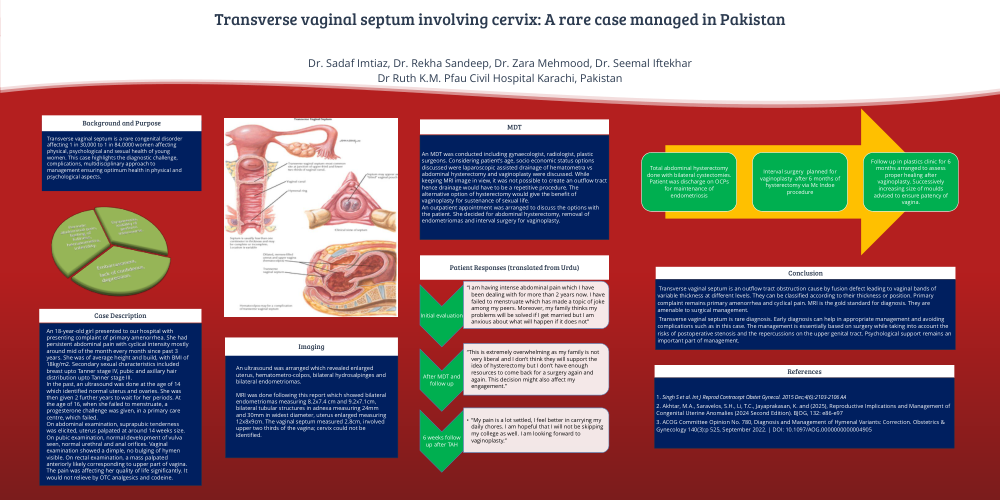
Transverse vaginal septum involving cervix: A rare case managed in Pakistan
Poster Presenter
Part of Topic
General gynaecology
Transverse vaginal septum involving cervix: A rare case managed in Pakistan
Dr. Sadaf Imtiaz, Dr. Rekha Sandeep, Dr. Zara Mehmood, Dr. Seemal Iftekhar
Dr Ruth K.M. Pfau Civil Hospital Karachi, Pakistan
Background and Purpose
Transverse vaginal septum is a rare congenital disorder
affecting 1 in 30,000 to 1 in 84,0000 women affecting
physical, psychological and sexual health of young
women. This case highlights the diagnostic challenge,
complications, multidisciplinary approach to
management ensuring optimum health in physical and
psychological aspects.
Case Description
An 18-year-old girl presented to our hospital with
presenting complaint of primary amenorrhea. She had
persistent abdominal pain with cyclical intensity mostly
around mid of the month every month since past 3
years. She was of average height and build, with BMI of
18kg/m2. Secondary sexual characteristics included
breast upto Tanner stage IV, pubic and axillary hair
distribution upto Tanner stage III.
In the past, an ultrasound was done at the age of 14
which identified normal uterus and ovaries. She was
then given 2 further years to wait for her periods. At
the age of 16, when she failed to menstruate, a
progesterone challenge was given, in a primary care
centre, which failed.
On abdominal examination, suprapubic tenderness
was elicited, uterus palpated at around 14 weeks size.
On pubic examination, normal development of vulva
seen, normal urethral and anal orifices. Vaginal
examination showed a dimple, no bulging of hymen
visible. On rectal examination, a mass palpated
anteriorly likely corresponding to upper part of vagina.
The pain was affecting her quality of life significantly. It
would not relieve by OTC analgesics and codeine.
Imaging
An ultrasound was arranged which revealed enlarged
uterus, hematometro-colpos, bilateral hydrosalpinges and
bilateral endometriomas.
MRI was done following this report which showed bilateral
endometriomas measuring 8.2x7.4 cm and 9.2x7.1cm,
bilateral tubular structures in adnexa measuring 24mm
and 30mm in widest diameter, uterus enlarged measuring
12x8x9cm. The vaginal septum measured 2.8cm, involved
upper two thirds of the vagina; cervix could not be
identified.
MDT
An MDT was conducted including gynaecologist, radiologist, plastic
surgeons. Considering patient’s age, socio economic status options
discussed were laparoscopic assisted drainage of hematometra vs
abdominal hysterectomy and vaginoplasty were discussed. While
keeping MRI image in view, it was not possible to create an outflow tract
hence drainage would have to be a repetitive procedure. The
alternative option of hysterectomy would give the benefit of
vaginoplasty for sustenance of sexual life.
An outpatient appointment was arranged to discuss the options with
the patient. She decided for abdominal hysterectomy, removal of
endometriomas and interval surgery for vaginoplasty.
Conclusion
Transverse vaginal septum is an outflow tract obstruction cause by fusion defect leading to vaginal bands of
variable thickness at different levels. They can be classified according to their thickness or position. Primary
complaint remains primary amenorrhea and cyclical pain. MRI is the gold standard for diagnosis. They are
amenable to surgical management.
Transverse vaginal septum is rare diagnosis. Early diagnosis can help in appropriate management and avoiding
complications such as in this case. The management is essentially based on surgery while taking into account the
risks of postoperative stenosis and the repercussions on the upper genital tract. Psychological support remains an
important part of management.
References
1. Singh S et al. Int J Reprod Contracept Obstet Gynecol. 2015 Dec;4(6):2103-2106 AA
2. Akhtar, M.A., Saravelos, S.H., Li, T.C., Jayaprakasan, K. and (2025), Reproductive Implications and Management of
Congenital Uterine Anomalies (2024 Second Edition). BJOG, 132: e86-e97
3. ACOG Committee Opinion No. 780, Diagnosis and Management of Hymenal Variants: Correction. Obstetrics &
Gynecology 140(3):p 525, September 2022. | DOI: 10.1097/AOG.0000000000004905