2,281 posters, 63 videos, 56 audios, 43 topics, 18 sessions, 6,174 authors, 2,323 institutions
ePostersLive by SciGen Technologies S.A. All rights reserved.
RCOG World Congress 2025
June 23 - 25, 2025 | ExCeL London, UK
EP.0616
Atypical presentation of Pseudo-Meigs Syndrome caused by Ovarian Fibrothecoma without pleural effusion
Poster Presenter
Authors
Affiliations
Part of Topic
General gynaecology
Atypical presentation of Pseudo-Meigs Syndrome caused by Ovarian Fibrothecoma without pleural effusion
Objective
Meigs syndrome (MS) is a rare condition characterised by a triad of benign ovarian fibroma, ascites and pleural effusion, occurring in approximately 1% of ovarian tumours. Pseudo-Meigs syndrome (P-MS), an even rarer entity, involves a triad of ascites, pleural effusion and pelvic or abdominal tumour other than fibroma, diagnosed post-operatively. Surgery is the primary treatment, with clinical resolution following tumour excision. Atypical presentation of P-MS without pleural effusion have been reported in a few cases often caused by uterine leiomyoma or ovarian metastasis from colon cancer only. We present a first case of atypical P-MS due to ovarian fibrothecoma associated with ascites, elevated Ca-125 without pleural effusion.
Case report
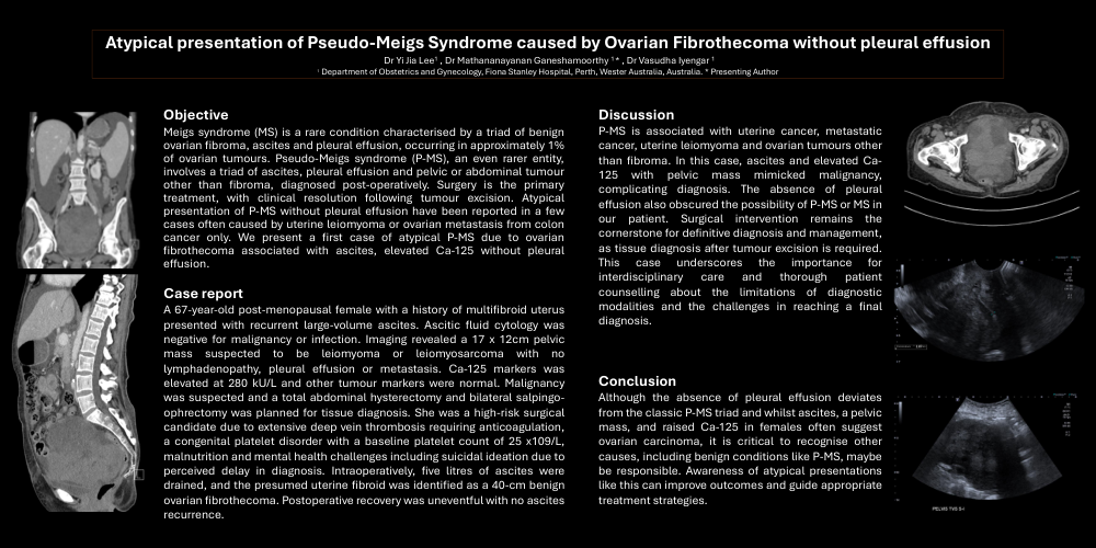
A 67-year-old post-menopausal female with a history of multifibroid uterus presented with recurrent large-volume ascites. Ascitic fluid cytology was negative for malignancy or infection. Imaging revealed a 17 x 12cm pelvic mass suspected to be leiomyoma or leiomyosarcoma with no lymphadenopathy, pleural effusion or metastasis. Ca-125 markers was elevated at 280 kU/L and other tumour markers were normal. Malignancy was suspected and a total abdominal hysterectomy and bilateral salpingo-ophrectomy was planned for tissue diagnosis.
She was a high-risk surgical candidate due to extensive deep vein thrombosis requiring anticoagulation, a congenital platelet disorder with a baseline platelet count of 25 x109/L, malnutrition and mental health challenges including suicidal ideation due to perceived delay in diagnosis. Intraoperatively, five litres of ascites were drained, and the presumed uterine fibroid was identified as a 40-cm benign ovarian fibrothecoma. Postoperative recovery was uneventful with no ascites recurrence.
Discussion
P-MS is associated with uterine cancer, metastatic cancer, uterine leiomyoma and ovarian tumours other than fibroma. In this case, ascites and elevated Ca-125 with pelvic mass mimicked malignancy, complicating diagnosis. The absence of pleural effusion also obscured the possibility of P-MS or MS in our patient. Surgical intervention remains the cornerstone for definitive diagnosis and management, as tissue diagnosis after tumour excision is required. This case underscores the importance for interdisciplinary care and thorough patient counselling about the limitations of diagnostic modalities and the challenges in reaching a final diagnosis.
Conclusion
Although the absence of pleural effusion deviates from the classic P-MS triad and whilst ascites, a pelvic mass and raised Ca-125 in females often suggest ovarian carcinoma, it is critical to recognise other causes, including benign conditions like P-MS, maybe be responsible. Awareness of atypical presentations like this can improve outcomes and guide appropriate treatment strategies.