2,281 posters, 63 videos, 56 audios, 43 topics, 18 sessions, 6,174 authors, 2,323 institutions
ePostersLive by SciGen Technologies S.A. All rights reserved.
RCOG World Congress 2025
June 23 - 25, 2025 | ExCeL London, UK
EP.0614
Leiomyoma arising from a rudimentary uterine horn in an MRKH patient
Poster Presenter
Part of Topic
General gynaecology
Leiomyoma arising from a rudimentary uterine horn in an MRKH patient
Dr. Elias Tsakos1, Dr. Emmanouil Xydias1, Dr. Vasileios Emmanouil2, Dr. Apostolos Ziogas3, Dr. Kanelina Bimpa1
1EmbryoClinic IVF, Thessaloniki, Greece. 2Democritus University of Thrace, School of Health Sciences, Faculty of Medicine, Alexandroupoli, Greece. 3University of Thessaly, School of Health Sciences, Faculty of Medicine, Larissa, Greece
Introduction
Mayer-Rokitansky-Küster-Hauser (MRKH) Syndrome is a rare condition associated with hypoplasia or even complete absence of the uterus. The lack of an intact reproductive tract frequently leads to Gynaecologists ignoring the risk of conventional uterine pathology occurring in these patients, thus potentially delaying diagnosis and leading to worse prognosis. The present report highlights this possibility, demonstrating a case of a large uterine fibroid originating from uterine remnants in an MRKH patient.
Case presentation
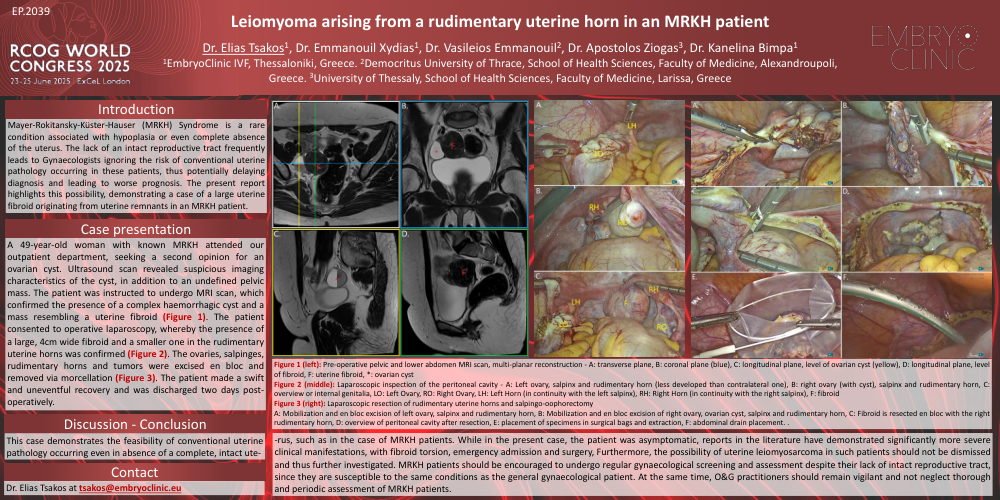
A 49-year-old woman with known MRKH attended our outpatient department, seeking a second opinion for an ovarian cyst. Ultrasound scan revealed suspicious imaging characteristics of the cyst, in addition to an undefined pelvic mass. The patient was instructed to undergo MRI scan, which confirmed the presence of a complex haemorrhagic cyst and a mass resembling a uterine fibroid (Figure 1). The patient consented to operative laparoscopy, whereby the presence of a large, 4cm wide fibroid and a smaller one in the rudimentary uterine horns was confirmed (Figure 2). The ovaries, salpinges, rudimentary horns and tumors were excised en bloc and removed via morcellation (Figure 3). The patient made a swift and uneventful recovery and was discharged two days post-operatively.
Discussion - Conclusion
This case demonstrates the feasibility of conventional uterine pathology occurring even in absence of a complete, intact uterus, such as in the case of MRKH patients. While in the present case, the patient was asymptomatic, reports in the literature have demonstrated significantly more severe clinical manifestations, with fibroid torsion, emergency admission and surgery, Furthermore, the possibility of uterine leiomyosarcoma in such patients should not be dismissed and thus further investigated. MRKH patients should be encouraged to undergo regular gynaecological screening and assessment despite their lack of intact reproductive tract, since they are susceptible to the same conditions as the general gynaecological patient. At the same time, O&G practitioners should remain vigilant and not neglect thorough and periodic assessment of MRKH patients.
Contact
Dr. Elias Tsakos at tsakos@embryoclinic.eu
Figure 1 (left): Pre-operative pelvic and lower abdomen MRI scan, multi-planar reconstruction - A: transverse plane, B: coronal plane (blue), C: longitudinal plane, level of ovarian cyst (yellow), D: longitudinal plane, level of fibroid, F: uterine fibroid, *: ovarian cyst
Figure 2 (middle): Laparoscopic inspection of the peritoneal cavity - A: Left ovary, salpinx and rudimentary horn (less developed than contralateral one), B: right ovary (with cyst), salpinx and rudimentary horn, C: overview or internal genitalia, LO: Left Ovary, RO: Right Ovary, LH: Left Horn (in continuity with the left salpinx), RH: Right Horn (in continuity with the right salpinx), F: fibroid
Figure 3 (right): Laparoscopic resection of rudimentary uterine horns and salpingo-oophorectomy
A: Mobilization and en bloc excision of left ovary, salpinx and rudimentary horn, B: Mobilization and en bloc excision of right ovary, ovarian cyst, salpinx and rudimentary horn, C: Fibroid is resected en bloc with the right rudimentary horn, D: overview of peritoneal cavity after resection, E: placement of specimens in surgical bags and extraction, F: abdominal drain placement. .