2,281 posters, 63 videos, 56 audios, 43 topics, 18 sessions, 6,174 authors, 2,323 institutions
ePostersLive by SciGen Technologies S.A. All rights reserved.
RCOG World Congress 2025
June 23 - 25, 2025 | ExCeL London, UK
EP.0609
Vulval leiomyoma mimicking Bartholin gland cyst: a case series
Poster Presenter
Part of Topic
General gynaecology
Vulval leiomyoma mimicking Bartholin gland cyst: a case series
Ramachandran Rathigashini1*, Shavindi Gunaratne1, WMPH Walisinghe1 and Rasika Herath1
1 Department of Obstetrics & Gynecology, Faculty of Medicine, University of Kelaniya, Sri Lanka
Objective
Vulval leiomyomas are rare benign smooth muscle tumours, accounting for 0.03% of all gynaecological tumours. The origin of these tumours is unclear. They are rarely observed before menarche and typically reduce following menopause. This suggests that hormones, specifically oestrogens and progesterone, may be involved in their growth. Tumour growth may also change significantly during pregnancy. Only 300 cases are reported so far in the literature. We report different presentations of two such cases.
Case Report
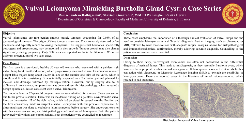
Our first case is a previously healthy 50-year-old woman who presented with a painless right vulval lump for 4 months’ duration, which progressively increased in size. Examination revealed a right labia majora lump about 3x2cm in size on the anterior one-third of the vulva, which is mobile and firm in consistency. It was initially suspected as a Bartholin cyst and planned for incision and drainage followed by marsupialization. However, during surgery due to its difference in consistency, lump excision was done and sent for histopathology, which revealed a benign spindle cell lesion consistent with a vulval leiomyoma.
Two months later, a 32-year-old pregnant woman was admitted for a repeat Caesarean section due to her previous section. There was an incidental finding of a painless, asymptomatic vulval lump on the anterior 1/3 of the right vulva, which had persisted for several months. Position and the firm consistency made us suspect a vulval leiomyoma with our previous experience. An ultrasound scan was done to exclude a leiomyosarcoma before surgery. She underwent excision with her caesarean section, and histopathology confirmed vulval leiomyoma. Both the patients recovered well without any complications. Both the patients were counselled on recurrence.
Discussion
Owing to their rarity, vulvovaginal leiomyomas are often not considered in the differential diagnosis of perineal lumps. This leads to misdiagnosis, as they resemble Bartholin cysts, which hampers the appropriate evaluation and management. If leiomyoma is suspected, it needs further evaluation with ultrasound or Magnetic Resonance Imaging (MRI) to exclude the possibility of leiomyosarcoma. There are reported cases in the literature of vulval leiomyosarcoma, which resulted in bad outcomes.
Conclusion
These cases emphasize the importance of a thorough clinical evaluation of vulval lumps and the need to consider leiomyomas as a differential diagnosis. Further imaging, such as ultrasound or MRI, followed by wide local excision with adequate surgical margins, allows for histopathological and immunohistochemical confirmation, thereby allowing accurate diagnosis. Counselling of the patients on recurrence is also essential.