2,281 posters, 63 videos, 56 audios, 43 topics, 18 sessions, 6,174 authors, 2,323 institutions
ePostersLive by SciGen Technologies S.A. All rights reserved.
RCOG World Congress 2025
June 23 - 25, 2025 | ExCeL London, UK
EP.0607
Missing Tubo Ovarian Complex - Looking Back 23 Years for Answers.
Poster Presenter
Authors
Affiliations
Part of Topic
General gynaecology
Missing Tubo Ovarian Complex - Looking Back 23 Years for Answers.
Presumed to be an antenatal torsion event, this case describes an unexpected intraoperative finding of tubo-ovarian absence and considerations should clinicians encounter this rare condition.
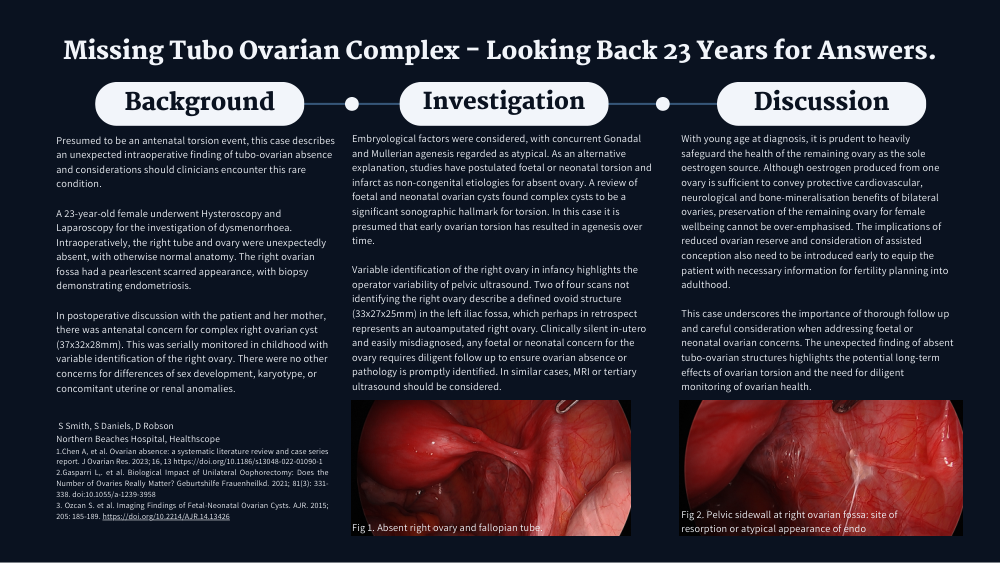
A 23-year-old female underwent Hysteroscopy and Laparoscopy for the investigation of dysmenorrhoea. Intraoperatively, the right tube and ovary were unexpectedly absent, with otherwise normal anatomy. The right ovarian fossa had a pearlescent scarred appearance, with biopsy demonstrating endometriosis. In postoperative discussion with the patient and her mother, there was antenatal concern for complex right ovarian cyst (37x32x28mm). This was serially monitored in childhood with variable identification of the right ovary. There were no other concerns for differences of sex development, karyotype, or concomitant uterine or renal anomalies.
Embryological factors were considered, with concurrent Gonadal and Mullerian agenesis regarded as atypical. As an alternative explanation, studies have postulated foetal or neonatal torsion and infarct as non-congenital etiologies for absent ovary. A review of foetal and neonatal ovarian cysts found complex cysts to be a significant sonographic hallmark for torsion [2].In this case it is presumed that early ovarian torsion has resulted in agenesis over time.
Variable identification of the right ovary in infancy highlights the operator variability of pelvic ultrasound. Two of four scans not identifying the right ovary describe a defined ovoid structure (33x27x25mm) in the left iliac fossa, which perhaps in retrospect represents an autoamputated right ovary. Clinically silent in-utero and easily misdiagnosed, any foetal or neonatal concern for the ovary requires diligent follow up to ensure ovarian absence or pathology is promptly identified. In similar cases, MRI or tertiary ultrasound should be considered.
With young age at diagnosis, it is prudent to heavily safeguard the health of the remaining ovary as the sole oestrogen source. Although oestrogen produced from one ovary is sufficient to convey protective cardiovascular, neurological and bone-mineralisation benefits of bilateral ovaries, preservation of the remaining ovary for female wellbeing cannot be over-emphasised. The implications of reduced ovarian reserve and consideration of assisted conception also need to be introduced early to equip the patient with necessary information for fertility planning into adulthood.
This case underscores the importance of thorough follow up and careful consideration when addressing foetal or neonatal ovarian concerns. The unexpected finding of absent tubo-ovarian structures highlights the potential long-term effects of ovarian torsion and the need for diligent monitoring of ovarian health.