2,281 posters, 63 videos, 56 audios, 43 topics, 18 sessions, 6,174 authors, 2,323 institutions
ePostersLive by SciGen Technologies S.A. All rights reserved.
RCOG World Congress 2025
June 23 - 25, 2025 | ExCeL London, UK
EP.0606
Parasitic Fibroid After Multiple Embolization in a Jehovah’s Witness Patient : Case Study
Poster Presenter
Authors
Amira Hassan, mohamed Elsawaf, Taner Kasapoglu, Samina Hameed, Bushra Saeed, Nabila Iram, Ghazi Ghazi
Affiliations
Part of Topic
General gynaecology
Parasitic Fibroid After Multiple Embolization in a Jehovah’s Witness Patient : Case Study
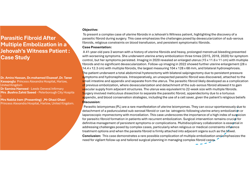
Objective: To present a complex case of uterine fibroids in a Jehovah’s Witness patient, highlighting the discovery of a parasitic fibroid during surgery. This case emphasizes the challenges posed by devascularization of sub-serous fibroids, religious constraints on blood transfusion, and persistent symptomatic fibroids.
Case Presentation: A 41-year-old para 3 woman with a history of uterine fibroids and heavy, prolonged menstrual bleeding presented with worsening symptoms. She underwent uterine artery embolization three times (2015, 2018, 2020) for symptom control, but her symptoms persisted. Imaging in 2020 revealed an enlarged uterus (15 x 11.6 x 11 cm) with multiple fibroids and no significant devascularization. Follow-up imaging in 2022 showed further uterine enlargement (28 x 14.4 x 12.3 cm) with multiple fibroids, the largest measuring 104 x 128 x 68 mm, and bilateral hydronephrosis.
The patient underwent a total abdominal hysterectomy with bilateral salpingectomy due to persistent pressure symptoms and hydronephrosis. Intraoperatively, an unexpected parasitic fibroid was discovered, attached to the small intestine and appendix and separate from the uterus. The parasitic fibroid likely developed as a complication of previous embolization, where devascularization and detachment of a the sub-serous fibroid allowed it to gain vascular supply from adjacent structures. The uterus was equivalent to 22-week size with multiple fibroids. Surgery involved meticulous dissection to separate the parasitic fibroid, appendectomy due to a tortuous appendix, and blood conservation strategies, including the use of a cell saver, given the patient’s religious beliefs.
Discussion: Parasitic leiomyomas (PL) are a rare manifestation of uterine leiomyomas. They can occur spontaneously due to detachment of a pedunculated sub-serosal fibroid or can be iatrogenic following uterine artery embolization or laparoscopic myomectomy with morcellation. This case underscores the importance of a high index of suspicion for parasitic fibroid formation in patients with recurrent embolization. Surgical intervention remains crucial for definitive management of persistent symptoms or complications. Multidisciplinary collaboration is essential in addressing challenges posed by complex cases, particularly when religious or medical constraints influence treatment options and when the parasite fibroid is firmly attached into adjacent organs such as the bowel.
Conclusion: This case demonstrates a rare possible complication of multiple embolization and emphasizes the need for vigilant follow-up and tailored surgical planning in managing complex fibroid cases.