2,281 posters, 63 videos, 56 audios, 43 topics, 18 sessions, 6,174 authors, 2,323 institutions
ePostersLive by SciGen Technologies S.A. All rights reserved.
RCOG World Congress 2025
June 23 - 25, 2025 | ExCeL London, UK
EP.0605
Atrophic Vesicovaginal fistula due to third degree Cystocele in an 89-year-old patient
Poster Presenter
Authors
Affiliations
Part of Topic
General gynaecology
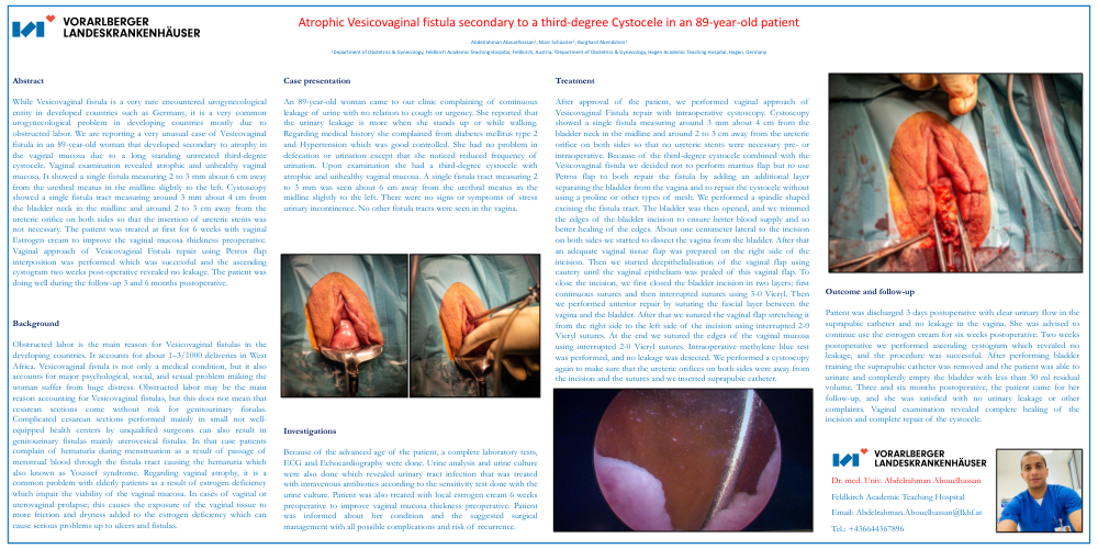
Atrophic Vesicovaginal fistula secondary to a third-degree Cystocele in an 89-year-old patient
Abdelrahman Abouelhassan1, Marc Schüssler2, Burghard Abendstein1
1Department of Obstetrics & Gynecology, Feldkirch Academic Teaching Hospital, Feldkirch, Austria, 2Department of Obstetrics & Gynecology, Hagen Academic Teaching Hospital, Hagen, Germany
Abstract
While Vesicovaginal fistula is a very rare encountered urogynecological
entity in developed countries such as Germany, it is a very common
urogynecological problem in developing countries mostly due to
obstructed labor. We are reporting a very unusual case of Vesicovaginal
fistula in an 89-year-old woman that developed secondary to atrophy in
the vaginal mucosa due to a long standing untreated third-degree
cystocele. Vaginal examination revealed atrophic and unhealthy vaginal
mucosa. It showed a single fistula measuring 2 to 3 mm about 6 cm away
from the urethral meatus in the midline slightly to the left. Cystoscopy
showed a single fistula tract measuring around 3 mm about 4 cm from
the bladder neck in the midline and around 2 to 3 cm away from the
ureteric orifice on both sides so that the insertion of ureteric stents was
not necessary. The patient was treated at first for 6 weeks with vaginal
Estrogen cream to improve the vaginal mucosa thickness preoperative.
Vaginal approach of Vesicovaginal Fistula repair using Petros flap
interposition was performed which was successful and the ascending
cystogram two weeks post-operative revealed no leakage. The patient was
doing well during the follow-up 3 and 6 months postoperative.
Background
Obstructed labor is the main reason for Vesicovaginal fistulas in the
developing countries. It accounts for about 1–3/1000 deliveries in West
Africa. Vesicovaginal fistula is not only a medical condition, but it also
accounts for major psychological, social, and sexual problem making the
woman suffer from huge distress. Obstructed labor may be the main
reason accounting for Vesicovaginal fistulas, but this does not mean that
cesarean sections come without risk for genitourinary fistulas.
Complicated cesarean sections performed mainly in small not well-
equipped health centers by unqualified surgeons can also result in
genitourinary fistulas mainly uterovesical fistulas. In that case patients
complain of hematuria during menstruation as a result of passage of
menstrual blood through the fistula tract causing the hematuria which
also known as Youssef syndrome. Regarding vaginal atrophy, it is a
common problem with elderly patients as a result of estrogen deficiency
which impair the viability of the vaginal mucosa. In cases of vaginal or
uterovaginal prolapse; this causes the exposure of the vaginal tissue to
more friction and dryness added to the estrogen deficiency which can
cause serious problems up to ulcers and fistulas.
Case presentation
An 89-year-old woman came to our clinic complaining of continuous
leakage of urine with no relation to cough or urgency. She reported that
the urinary leakage is more when she stands up or while walking.
Regarding medical history she complained from diabetes mellitus type 2
and Hypertension which was good controlled. She had no problem in
defecation or urination except that she noticed reduced frequency of
urination. Upon examination she had a third-degree cystocele with
atrophic and unhealthy vaginal mucosa. A single fistula tract measuring 2
to 3 mm was seen about 6 cm away from the urethral meatus in the
midline slightly to the left. There were no signs or symptoms of stress
urinary incontinence. No other fistula tracts were seen in the vagina.
Investigations
Because of the advanced age of the patient, a complete laboratory tests,
ECG and Echocardiography were done. Urine analysis and urine culture
were also done which revealed urinary tract infection that was treated
with intravenous antibiotics according to the sensitivity test done with the
urine culture. Patient was also treated with local estrogen cream 6 weeks
preoperative to improve vaginal mucosa thickness preoperative. Patient
was informed about her condition and the suggested surgical
management with all possible complications and risk of recurrence.
Treatment
After approval of the patient, we performed vaginal approach of
Vesicovaginal Fistula repair with intraoperative cystoscopy. Cystoscopy
showed a single fistula measuring around 3 mm about 4 cm from the
bladder neck in the midline and around 2 to 3 cm away from the ureteric
orifice on both sides so that no ureteric stents were necessary pre- or
intraoperative. Because of the third-degree cystocele combined with the
Vesicovaginal fistula we decided not to perform martius flap but to use
Petros flap to both repair the fistula by adding an additional layer
separating the bladder from the vagina and to repair the cystocele without
using a proline or other types of mesh. We performed a spindle shaped
excising the fistula tract. The bladder was then opened, and we trimmed
the edges of the bladder incision to ensure better blood supply and so
better healing of the edges. About one centimeter lateral to the incision
on both sides we started to dissect the vagina from the bladder. After that
an adequate vaginal tissue flap was prepared on the right side of the
incision. Then we started deepithelialisation of the vaginal flap using
cautery until the vaginal epithelium was pealed of this vaginal flap. To
close the incision, we first closed the bladder incision in two layers; first
continuous sutures and then interrupted sutures using 3-0 Vicryl. Then
we performed anterior repair by suturing the fascial layer between the
vagina and the bladder. After that we sutured the vaginal flap stretching it
from the right side to the left side of the incision using interrupted 2-0
Vicryl sutures. At the end we sutured the edges of the vaginal mucosa
using interrupted 2-0 Vicryl sutures. Intraoperative methylene blue test
was performed, and no leakage was detected. We performed a cystoscopy
again to make sure that the ureteric orifices on both sides were away from
the incision and the sutures and we inserted suprapubic catheter.
Outcome and follow-up
Patient was discharged 3 days postoperative with clear urinary flow in the
suprapubic catheter and no leakage in the vagina. She was advised to
continue use the estrogen cream for six weeks postoperative. Two weeks
postoperative we performed ascending cystogram which revealed no
leakage, and the procedure was successful. After performing bladder
training the suprapubic catheter was removed and the patient was able to
urinate and completely empty the bladder with less than 30 ml residual
volume. Three and six months postoperative, the patient came for her
follow-up, and she was satisfied with no urinary leakage or other
complaints. Vaginal examination revealed complete healing of the
incision and complete repair of the cystocele.