2,281 posters, 63 videos, 56 audios, 43 topics, 18 sessions, 6,174 authors, 2,323 institutions
ePostersLive by SciGen Technologies S.A. All rights reserved.
RCOG World Congress 2025
June 23 - 25, 2025 | ExCeL London, UK
EP.0590
A large cystic uterine mass: a diagnostic dilemma
Poster Presenter
Authors
Affiliations
Part of Topic
General gynaecology
A Large CysticUterine Mass: A Diagnostic Dilemma
Introduction
Uterine leiomyomas are a common benign proliferation of uterine smooth muscle commonly seen in women of reproductive age. They are a common finding during pregnancy and can result in complications such as preterm labour, growth restriction and pain secondary to fibroid degeneration. The most common type of fibroid degeneration in pregnancy is red degeneration which is a subtype of haemorrhagic infarction of leiomyomas. The exact mechanism of red degeneration in pregnancy is not fully understood, however it is thought to be a result of rapid growth of the fibroid in early pregnancy resulting in inadequate blood supply.
We present a case of a primigravida presenting with a large fibroid which has grown exponentially in the second trimester with an intraoperative appearance of an adenomyoma.
Case presentation
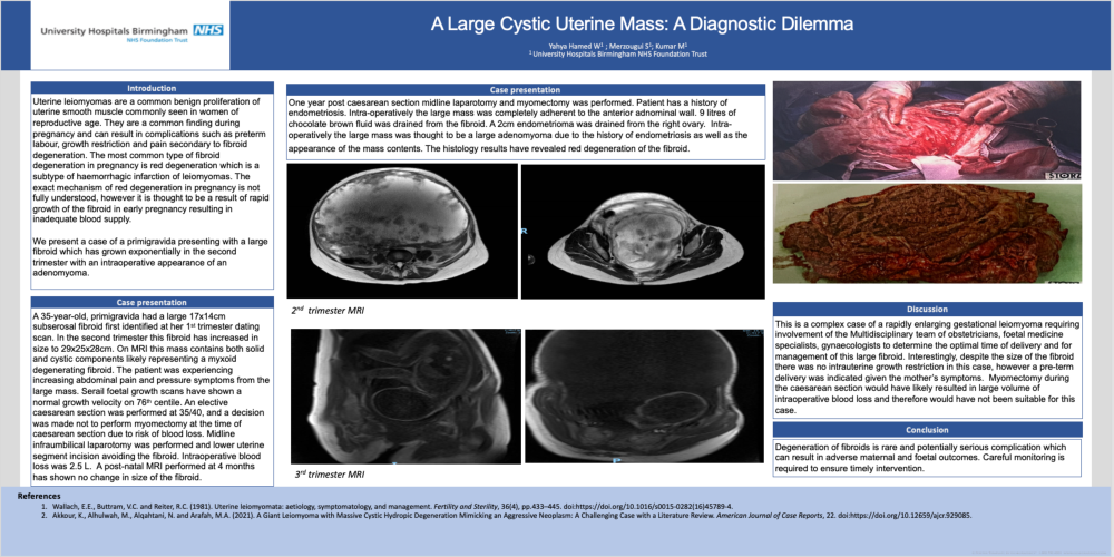
A 35-year-old, primigravida had a large 17x14cm subserosal fibroid first identified at her 1st trimester dating scan. In the second trimester this fibroid has increased in size to 29x25x28cm. On MRI this mass contains both solid and cystic components likely representing a myxoid degenerating fibroid. The patient was experiencing increasing abdominal pain and pressure symptoms from the large mass. Serail foetal growth scans have shown a normal growth velocity on 76th centile. An elective caesarean section was performed at 35/40, and a decision was made not to perform myomectomy at the time of caesarean section due to risk of blood loss. Midline infraumbilical laparotomy was performed and lower uterine segment incision avoiding the fibroid. Intraoperative blood loss was 2.5 L. A post-natal MRI performed at 4 months has shown no change in size of the fibroid.
One year post caesarean section midline laparotomy and myomectomy was performed. Patient has a history of endometriosis. Intra-operatively the large mass was completely adherent to the anterior adnominal wall. 9 litres of chocolate brown fluid was drained from the fibroid. A 2cm endometrioma was drained from the right ovary. Intra-operatively the large mass was thought to be a large adenomyoma due to the history of endometriosis as well as the appearance of the mass contents. The histology results have revealed red degeneration of the fibroid.
Discussion
This is a complex case of a rapidly enlarging gestational leiomyoma requiring involvement of the Multidisciplinary team of obstetricians, foetal medicine specialists, gynaecologists to determine the optimal time of delivery and for management of this large fibroid. Interestingly, despite the size of the fibroid there was no intrauterine growth restriction in this case, however a pre-term delivery was indicated given the mother’s symptoms. Myomectomy during the caesarean section would have likely resulted in large volume of intraoperative blood loss and therefore would have not been suitable for this case.
Conclusion
Degeneration of fibroids is rare and potentially serious complication which can result in adverse maternal and foetal outcomes. Careful monitoring is required to ensure timely intervention.