2,281 posters, 63 videos, 56 audios, 43 topics, 18 sessions, 6,174 authors, 2,323 institutions
ePostersLive by SciGen Technologies S.A. All rights reserved.
RCOG World Congress 2025
June 23 - 25, 2025 | ExCeL London, UK
EP.0586
Management of Giant Prolapsed Submucous Leiomyoma: A Surgical Challenge for Gynecologists
Poster Presenter
Authors
Affiliations
Part of Topic
General gynaecology
Management of Giant Prolapsed Submucous Leiomyoma: A Surgical Challenge for Gynecologists
Objective:
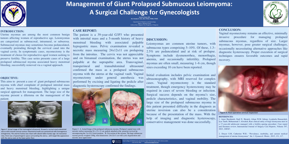
This paper aims to present a case of giant prolapsed submucous myoma with chief complaint of prolapsed introital mass and heavy menstrual bleeding, highlighting a unique surgical approach for management. The large size of the myoma present a dilemma on the management of the patient.
Case Report:
The patient is a 39-year-old G3P3 who presented with intoital mass and a 5-month history of heavy menstrual bleeding with associated palpable hypogastric mass. Pelvic examination revealed a necrotic mass measuring 24x12x11 cm prolapsing from the introitus. The cervix was not appreciable and on bimanual examination, the uterus was not palpable at the suprapubic area. Transvaginal, transperineal and transabdominal ultrasound confirmed the mass as a prolapsed submucous myoma with the uterus at the vaginal vault. Vaginal myomectomy under general anesthesia was performed by excising and ligating the pedicle after diagnostic hysteroscopy confirmed the findings.
Discussion:
Leiomyomas are common uterine tumors, with submucous types comprising 5–10%. Of these, 1.3–2.5% are pedunculated and at risk of prolapse. Symptoms include dysmenorrhea, menorrhagia, anemia, and occasionally infertility. Prolapsed myomas are often small, measuring 1–6 cm, though sizes exceeding 10 cm have been reported.
Initial evaluation includes pelvic examination and ultrasonography, with MRI reserved for complex cases. Vaginal myomectomy is the standard treatment, though emergency hysterectomy may be required in cases of severe bleeding or infection. Surgical success depends on the myoma’s size, pedicle characteristics, and vaginal mobility. The large size of the prolapsed submucous myoma in this patient presented difficulty in the diagnosis as uterine inversion can also be a consideration because of the presentation of the mass. With the help of imaging and diagnostic hysteroscopy, conservative management was done successfully.
Conclusion:
Vaginal myomectomy remains an effective, minimally invasive procedure for managing prolapsed submucous myomas, regardless of size. Larger myomas, however, pose greater surgical challenges, occasionally necessitating alternative approaches like diagnostic hysteroscopy. Proper execution of surgical techniques ensures favorable outcomes and rapid recovery.