2,281 posters, 63 videos, 56 audios, 43 topics, 18 sessions, 6,174 authors, 2,323 institutions
ePostersLive by SciGen Technologies S.A. All rights reserved.
RCOG World Congress 2025
June 23 - 25, 2025 | ExCeL London, UK
EP.0584
Placental site nodule (PSN)- An uncommon diagnosis with a common presentation
Poster Presenter
Authors
Affiliations
Part of Topic
General gynaecology
PLACENTAL SITE NODULE: An Uncommon Diagnosis With A Common Presentation
Megha Batra, Ashok Mohanty, Shiranthika Wickramasuriya
INTRODUCTION
Placental site nodule (PSN) is a rare, benign lesion of the intermediate trophoblast.
Thought to represent remnants from the previous pregnancy that have failed to involute completely.
Usually occurs in the reproductive age group.
Often the time interval between pregnancy and diagnosis of PSN can be several years.
Majority involve the endometrium, PSN can occasionally be seen in the cervix and rarely in the Fallopian tube and ovary
It represents the benign counterpart of epithelioid trophoblastic tumour (ETT).
Atypical placental site nodule (APSN) is a term assigned to placental site nodules with significant nuclear atypia or borderline proliferation index.
Atypical PSN can potentially progress to placental site trophoblastic tumour (PSTT) and epithelial trophoblastic tumour.
CLINICAL FEATURES
Asymptomatic
Or usually present as heavy menstrual bleeding (HMB) or intermenstrual irregular bleeding or abnormal cervical smear.
Infertility or recurrent pregnancy loss is a rare mode of presentation in PSN.
CASE PRESENTATION
34-year lady, Para 2, with a history of polycystic ovarian syndrome, presenting with irregular, heavy menstrual bleeding and left iliac fossa pain, for the last 4 years since her childbirth.
Previous both full term normal vaginal deliveries with no complications.
No relief with non hormonal and hormonal treatment options in the form of contraceptive pills.
General physical examination, routine gynaecological checkup, cervical smear normal.
Serum beta HCG negative with Haemoglobin of 136.
INVESTIGATIONS
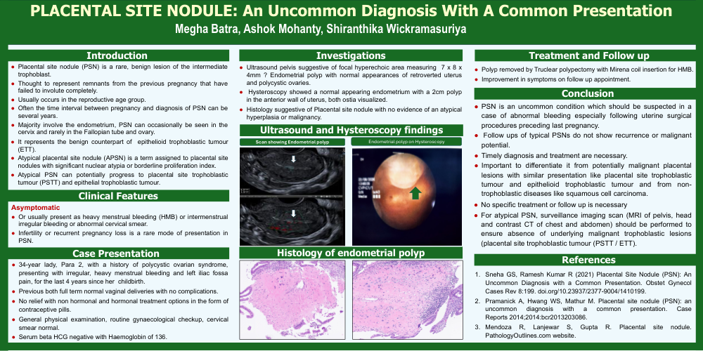
Ultrasound pelvis suggestive of focal hyperechoic area measuring 7 x 8 x 4mm ? Endometrial polyp with normal appearances of retroverted uterus and polycystic ovaries.
Hysteroscopy showed a normal appearing endometrium with a 2cm polyp in the anterior wall of uterus, both ostia visualized.
Histology suggestive of Placental site nodule with no evidence of an atypical hyperplasia or malignancy.
TREATMENT AND FOLLOW UP
Polyp removed by Truclear polypectomy with Mirena coil insertion for HMB.
Improvement in symptoms on follow up appointment
CONCLUSION
PSN is an uncommon condition which should be suspected in a case of abnormal bleeding especially following uterine surgical procedures preceding last pregnancy.
Follow ups of typical PSNs do not show recurrence or malignant potential.
Timely diagnosis and treatment are necessary.
Important to differentiate it from potentially malignant placental lesions with similar presentation like placental site trophoblastic tumour and epithelioid trophoblastic tumour and from non- trophoblastic diseases like squamous cell carcinoma.
No specific treatment or follow up is necessary
For atypical PSN, surveillance imaging scan (MRI of pelvis, head and contrast CT of chest and abdomen) should be performed to ensure absence of underlying malignant trophoblastic lesions (placental site trophoblastic tumour (PSTT / ETT).