2,281 posters, 63 videos, 56 audios, 43 topics, 18 sessions, 6,174 authors, 2,323 institutions
ePostersLive by SciGen Technologies S.A. All rights reserved.
RCOG World Congress 2025
June 23 - 25, 2025 | ExCeL London, UK
EP.0582
Progressive pilonidal disease of the peri-clitoris: a case report, review and discussion
Poster Presenter
Authors
Affiliations
Part of Topic
General gynaecology
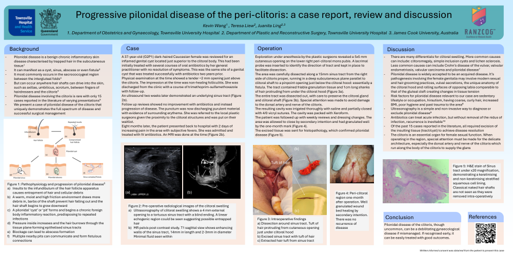
Figure 1: Pathophysiology and progression of pilonidal disease6
A 37-year-old (G2P1) dark-haired Caucasian female was reviewed for an inflamed genital cyst located just superior to the clitoral body. This had been initially treated with several courses of oral antibiotics by her general practitioner with no resolution of symptoms. This was the same site of a genital cyst that was treated successfully with antibiotics two years prior.
Physical examination at the time showed a tender ~2 mm opening just above the clitoris. The impression at the time was non-healing folliculitis. She was discharged from the clinic with a course of trimethoprim-sulfamethoxazole with follow-up.
An ultrasound two weeks later, demonstrated a sinus tract (Figure 2a).
The follow up reviews showed no improvement with antibiotics and instead progression of disease. The punctum was now discharging purulent material with evidence of surrounding erythema. She was referred to the local plastic surgeons given the proximity to the clitoral structures and was put on their waitlist.
Eight months later, the patient presented back to hospital with 2 days of increasing pain in the area with subjective fevers. She was admitted and treated with IV antibiotics. An MRI was done at the time (Figure 2b).
Figure 2: Pre-operative radiological images of the clitoral swelling
Exploration under anesthesia by the plastic surgeons revealed a 5x5 mm cutaneous opening on the lower right peri-clitoral mons pubis. A lacrimal probe was inserted to identify the direction of tract and kept in place to facilitate dissection.
The area was carefully dissected along a 15mm sinus tract from the right side of clitoris proper, running in a deep subcutaneous plane parallel to clitoral shaft to a pinpoint opening just above the clitoral hood: essentially a fistula. The tract contained friable granulation tissue and 1cm long shanks of hair protruding from under the clitoral hood (Figure 3a).
The entire tract was dissected out, with care to preserve the clitoral gland and clitoral shaft (Figure 3b). Special attention was made to avoid damage to the dorsal artery and nerve of the clitoris.
The resulting cavity was irrigated thoroughly with saline and partially closed with 4/0 vicryl sutures. The cavity was packed with Xeroform.
The patient was followed up with weekly reviews and dressing changes. The area was allowed to close by secondary intention and had granulated well by the 1-month mark (Figure 4).
The excised tissue was sent for histopathology which confirmed pilonidal disease (Figure 5).
Figure 3: Intraoperative findings
a) Dissection around sinus tract. Tuft of hair protruding from cutaneous opening just under clitoral hood
b) Excised sinus tract with tuft of hair
c) Extracted hair tuft from sinus tract
Figure 4: Peri-clitoral region 1-month after operation. Well granulated wound bed healing by secondary intention. There was no recurrence of disease
Figure 5: H&E stain of Sinus tract under x20 magnification, demonstrating a keratinising and non-keratinising stratified squamous cell lining. Classical naked hair shafts are not seen as they were removed intra-operatively
Pilonidal disease of the clitoris, though uncommon, can be a debilitating gynaecological disease if mismanaged. If recognised early, it can be easily treated with good outcomes.