2,281 posters, 63 videos, 56 audios, 43 topics, 18 sessions, 6,174 authors, 2,323 institutions
ePostersLive by SciGen Technologies S.A. All rights reserved.
RCOG World Congress 2025
June 23 - 25, 2025 | ExCeL London, UK
EP.0573
A rare case of isolated primary leiomyoma discovered within an ovarian endometrioma
Poster Presenter
Part of Topic
General gynaecology
Title: A Rare Case of Isolated Primary Leiomyoma discovered within an Ovarian Endometrioma: Case Report
Case Report:
A 36-year-old nulliparous woman was admitted to the Singapore General Hospital with a 1 day history of acute lower abdominal pain not associated with her menstrual cycle or vaginal bleeding. Physical examination revealed a palpable pelvic mass up to the level of the umbilicus and an acute abdomen presentation with generalized tenderness and abdominal guarding over the suprapubic and left lower abdominal regions. The rest of her clinical parameters were within normal ranges.
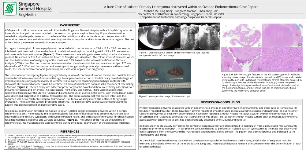
An urgent transvaginal ultrasonography was conducted which demonstrated a 13.6 x 11.8 x 13.0 centimetres lobulated cystic mass with low level echoes in the left adnexal region containing a 6.0 x 3.3 x 3.7 centimetres solid area with tiny cystic spaces (Figure 1). There were also some echogenic areas with posterior shadowing present. No ascites or free fluid within the Pouch of Douglas was visualized. The colour score of this mass was 3 and the likelihood ratio of malignancy of this mass was 6.9% based on the International Ovarian Tumour Analysis (IOTA) score. The uterus was otherwise normal on the ultrasound. Her serum cancer antigen 125 was elevated at 46.6 U/ml, and her serum carcinoembryonic antigen and alpha-fetal protein were within normal limits. The serum human chorionic gonadotrophin levels were negative for pregnancy.
She underwent an emergency laparotomy cystectomy in view of concerns of ovarian torsion and possible loss of ovarian function in a woman of reproductive age. Intraoperative inspection of the left ovary revealed a large left ovarian cyst measuring approximately 14-centimetres in diameter containing hemorrhagic contents with chocolate-like fluid and another small 6.5-centimetres area of whitish firm nodular solid component resembling a fibroma (Figure 2). The left ovary was adherent posteriorly to the bowel and there were flimsy adhesions over the anterior uterus and left ovary. The contralateral right ovary was normal. There were multiple small subserosal fibroids over the uterine fundus and a small amount of ascites in the pelvis. Both the fallopian tubes were distended, suggestive of bilateral hydrosalpinges. The entire ovarian cyst was excised intact and the specimen was delivered for histological examination. Peritoneal washings were also obtained for cytologic evaluation. The rest of the surgery proceeded smoothly. The postoperative course was uneventful and the patient was discharged well on postoperative day 2.
Histological examination revealed the nodule to be an isolated benign ovarian leiomyoma within a benign endometriotic cyst. Histologically, the nodule displayed fascicles of bland spindled smooth muscle cells with eosinophilic and fibrillary cytoplasm, with ovoid elongated nuclei, and with areas of interstitial fibrohyalinisation, focal haemorrhage, oedema, and variable cellularity (Figure 3). The surface of the nodule showed foci of endometriosis. No malignant cells were identified during cytological examination of the peritoneal washings.
Discussion/Conclusion:
Primary ovarian leiomyoma associated with an endometriotic cyst is an extremely rare finding and only one other case by Tomas et al [1] has been reported thus far. There have been several reports of smooth muscle metaplasia within ovarian endometriosis [2], but no other cases of leiomyomas arising from endometriosis have been reported. Smooth muscle metaplasia within ovarian endometriosis is not uncommon and Fukaunaga estimates that its prevalence was about 18% [2]. Other smooth muscle tumors such as ovarian adenomyoma associated with endometriotic cyst has been previously described by McDougal and Roth [3].
Radical surgeries are usually performed for complete excision as they are often difficult to distinguish from a pelvic solid mass and rarely diagnosed prior to operation [4]. In our present case, we decided to perform an isolated ovarian cystectomy as the mass was clearly and easily separable from the ovary and the macroscopic appearances looked benign. The patient was also nulliparous and belonged to the reproductive age group.
As the clinical manifestation may vary with no consistent sonographic or biochemical distinctions, a high index of suspicion must be exercised particularly in women of the reproductive age group. Histological diagnosis remains the cornerstone for the determination of such unusual pathology.
References: