2,281 posters, 63 videos, 56 audios, 43 topics, 18 sessions, 6,174 authors, 2,323 institutions
ePostersLive by SciGen Technologies S.A. All rights reserved.
RCOG World Congress 2025
June 23 - 25, 2025 | ExCeL London, UK
EP.0567
A very rare Case of Spontaneous rupture of degenerated uterine fibroid
Poster Presenter
Part of Topic
General gynaecology
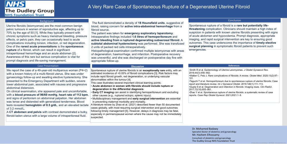
A Very Rare Case of Spontaneous Rupture of a Degenerated Uterine Fibroid
Background
Uterine fibroids (leiomyomas) are the most common benign
pelvic tumours in women of reproductive age, affecting up to
70% by the age of 50 [1]. While they typically present with
chronic symptoms such as heavy menstrual bleeding, pressure
effects, or pelvic pain, they can occasionally lead to acute
complications including torsion, infection, or red degeneration.
One of the rarest acute presentations is the spontaneous
rupture of a fibroid, which can result in significant
intraperitoneal hemorrhage and an acute abdomen [2].
Awareness of this rare but serious complication is vital for
prompt diagnosis and life-saving management.
Case Presentation
We report the case of a 49-year-old multiparous woman (P4+2)
with a known history of a multi-fibroid uterus. She was under
gynaecology follow-up and awaiting elective hysterectomy. She
presented to the Emergency Department with sudden, severe
lower abdominal pain, associated with nausea and progressive
abdominal distension.
On clinical examination, she appeared pale and uncomfortable,
with a blood pressure of 96/60 mmHg, heart rate of 112 bpm,
and signs of peritonism on abdominal palpation. Her abdomen
was tense and distended with generalized tenderness. Blood
tests revealed hemoglobin of 8.5 g/dL, and an elevated lactate
of 3.2 mmol/L.
A CT abdomen and pelvis with contrast demonstrated a bulky,
fibroid-laden uterus with a large volume of intraperitoneal fluid.
The fluid demonstrated a density of 18 Hounsfield units, suggestive of
blood, raising concern for active intra-abdominal hemorrhage from a
fibroid source.
The patient was taken for emergency exploratory laparotomy.
Intraoperative findings included 1.6 litres of hemoperitoneum and
active bleeding from a ruptured degenerating subserosal fibroid. A
subtotal abdominal hysterectomy was performed. She was transfused
2 units of packed red cells intraoperatively.
Histopathological examination confirmed multiple leiomyomas with areas
of degeneration, haemorrhage, and infarction. Postoperative recovery
was uneventful, and she was discharged on postoperative day five with
appropriate follow-up.
Discussion
Spontaneous rupture of uterine fibroids is an exceptionally rare entity, with an
estimated incidence of <0.05% of fibroid complications [3]. Risk factors may
include rapid fibroid growth, red degeneration, or underlying vascular
abnormalities within the tumour [4].
This case illustrates several important clinical learning points:
• Acute abdomen in women with fibroids should include rupture or
degeneration in the differential diagnosis.
• Early CT imaging can assist in identifying hemoperitoneum and excluding
other causes (e.g., ruptured ectopic, splenic injury).
• Multidisciplinary management and early surgical intervention are essential
in preventing maternal morbidity and mortality.
A literature review by Zhao et al. (2021) described fewer than 50 documented
cases globally, with most requiring surgical intervention and good outcomes
following timely management [5]. However, delays in diagnosis may be fatal,
especially in perimenopausal women where the cause may not be immediately
suspected.
conclusion
Spontaneous rupture of a fibroid is a rare but potentially life-
threatening complication. Clinicians should maintain a high index of
suspicion in patients with known uterine fibroids presenting with signs
of acute abdomen and hypovolemia. Prompt diagnosis, appropriate
imaging, and rapid surgical intervention are key to ensuring good
outcomes. This case underscores the importance of timely elective
surgical planning in symptomatic fibroid patients to prevent such
emergencies.
References
•Smith R et al. Epidemiology of Uterine Leiomyomas. J Obstet Gynaecol Res.
2018;44(3):456–462.
•Walker C, Pelz J. Rare complications of fibroids: A review. Obstet Med. 2020;13(2):97–
101.
•Taguchi T et al. Hemoperitoneum due to spontaneous rupture of uterine fibroids: Case
report and literature review. Int J Gynaecol Obstet. 2019;146(1):111–113.
•Gupta S et al. Degeneration and infarction in fibroids: Imaging clues. Clin Radiol.
2017;72(10):855–861.
•Zhao Y et al. Spontaneous rupture of uterine fibroids: a systematic review of case
reports. Case Rep Obstet Gynecol. 2021;2021:1–8.
Dr. Mohamed Badawy
Specialist Doctor of obstetrics and gynaecology
Mr. Hashem Elhossamy
Consultant of obstetrics and gynaecology
The Dudley Group NHS Foundation Trust