2,281 posters, 63 videos, 56 audios, 43 topics, 18 sessions, 6,174 authors, 2,323 institutions
ePostersLive by SciGen Technologies S.A. All rights reserved.
RCOG World Congress 2025
June 23 - 25, 2025 | ExCeL London, UK
EP.0516
Investigating the hormone and prostaglandin receptor profiles of the human myometrium.
Poster Presenter
Authors
Lorna Salvini, Alison Maclean, Christopher Hill, Dharani Hapangama
Affiliations
Part of Topic
General gynaecology
Comparing hormone receptor expression in the inner and outer layers of the human myometrium during labour and across the menstrual cycle.
Lorna Salvini, Alison Maclean, Christopher Hill, Eva Caamaño Gutierrez, Parveen Sharma & Dharani Hapangama
Department of Women's and Children's Health, Institute of Life Course and Medical Sciences, University of Liverpool, UK L8 7SS
Background & Hypothesis:
The myometrium, the uterine muscular wall, plays an essential role in menstruation and parturition by generating and coordinating contractions. It has two distinct layers: the inner junctional zone and the outer longitudinal layer, hypothesised to have different embryonic origins.
Hormone-responsive myometrial pathologies like leiomyoma and adenomyosis, often cause debilitating symptoms e.g. chronic pelvic pain and heavy menstrual bleeding, impact quality of life and affect one in three women of reproductive age. The hormone receptor profiles of the myometrial layers have not yet been fully characterised. The cellular composition and structure of the non-pregnant myometrium is poorly understood, with limited research on myometrial layers. Therefore, it is hypothesised that… The two myometrial layers have distinct hormone receptor profiles, influencing uterine function and pathologies.
Methods:
Immunohistochemistry was used to detect the presence of oestrogen receptor (ERa), progesterone receptor (PR) and androgen receptor (AR) in full thickness myometrial samples obtained during the proliferative phase (PP, n=10), secretory phase (SP, n=10), and in term, non-labouring pregnancy (TP, n=10). Staining was quantified using StarDist software in ImageJ and graphed in R studio. A Mann-Whitney U test was used to compare expression between the inner and outer myometrial layers. Significance was considered for p < 0.05.
Results:
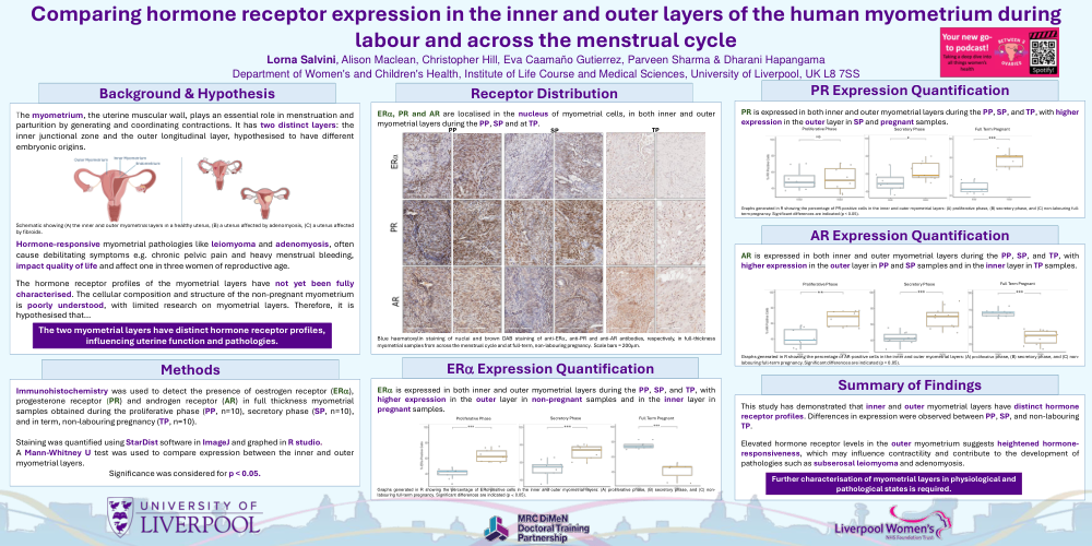
ERa, PR and AR are localised i the nucleus of myometrial cells, in both inner and outer myometrial layers during hte PP, SP and at TP.
ERa is expressed in both inner and outer myometrial layers during the PP, SP, and TP, with higher expression in the outer layer in non-pregnant samples and in the inner layer in pregnant samples.
PR is expressed in both inner and outer myometrial layers during the PP, SP, and TP, with higher expression in the outer layer in SP and pregnant samples.
AR is expressed in both inner and outer myometrial layers during the PP, SP, and TP, with higher expression in the outer layer in PP and SP samples and in the inner layer in TP samples.
Summary of Findings:
This study has demonstrated that inner and outer myometrial layers have distinct hormone receptor profiles. Differences in expression were observed between PP, SP, and non-labouring TP. Elevated hormone receptor levels in the outer myometrium suggests heightened hormoneresponsiveness, which may influence contractility and contribute to the development of pathologies such as subserosal leiomyoma and adenomyosis.
Further characterisation of myometrial layers in physiological and patholigical states is required.