2,281 posters, 63 videos, 56 audios, 43 topics, 18 sessions, 6,174 authors, 2,323 institutions
ePostersLive by SciGen Technologies S.A. All rights reserved.
RCOG World Congress 2025
June 23 - 25, 2025 | ExCeL London, UK
EP.0430
When CTG leads to prenatal diagnosis: two cases of fetal intestinal volvulus
Poster Presenter
Part of Topic
Fetal medicine
When CTG leads to prenatal diagnosis: two cases of fetal intestinal volvulus
Nina Navakumar1, Alice Pastides1, Lucius Baltoaica1, ManHo Kwok1, Susana Pereira1, Elena Greco1,2.
1Royal London Hospital, Barts Health NHS Trust; 2 Women’s Health Research Unit, Wolfson Institute of Population Health, Queen Mary University London.
nina.navakumar@nhs.net
Alice.pastides@nhs.net, Lucius.baltoaica@nhs.net, manho.kwok2@nhs.net, Susana.pereira@nhs.net, e.greco@qmul.ac.uk
Abstract:
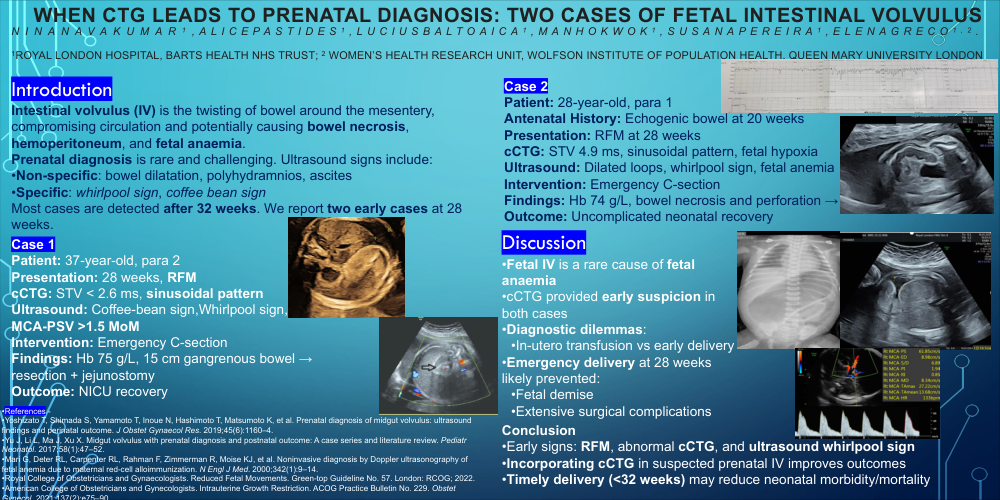
Intestinal volvulus (IV) is the twisting of the intestine around the mesentery, compromising venous return and leading to bowel dilatation, necrosis, and potentially fatal complications such as hemoperitoneum, fetal anaemia, and death. Prenatally, IV is a rare but critical diagnosis requiring timely intervention. Ultrasound signs of IV include non-specific features like bowel dilatation, polyhydramnios, and ascites, and specific features such as the whirlpool or coffee bean sign. Most reported cases are diagnosed after 32 weeks by ultrasound, with close surveillance and delivery by 34 weeks often recommended. We report two cases of IV with severe fetal anaemia diagnosed at 28 weeks’ gestation following reduced fetal movements (RFM) and abnormal computerised CTG (cCTG), detailing the diagnostic approach, emergency management, and neonatal outcomes.
Case 1: A 37-year-old para 2 presented at 28 weeks with first-episode RFM. cCTG showed reduced short-term variation (STV < 2.6 ms) and a sinusoidal pattern indicative of fetal anaemia. Ultrasound confirmed the whirlpool sign and MCA-PSV >1.5 MoM, suggestive of IV and fetal anaemia. Emergency caesarean section was performed, revealing neonatal anaemia (Hb 75 g/L) and IV with 15 cm of gangrenous bowel. The neonate underwent resection and jejunostomy, recovering well in NICU.
Case 2: A 28-year-old para 1 with known fetal echogenic bowel at 20 weeks presented with RFM at 28 weeks. Abnormal cCTG (STV 4.9 ms and sinusoidal pattern) prompted urgent ultrasound, which showed dilated bowel loops, whirlpool sign, and fetal anaemia. Emergency caesarean delivery revealed severe neonatal anaemia (Hb 74 g/L) and IV with necrosis and perforation. Surgical resection and primary anastomosis were performed, with an uncomplicated neonatal recovery.
Discussion: Midgut volvulus is a rare cause of fetal anaemia, identifiable by pathognomonic ultrasound signs. While fetal anaemia is a known complication of IV, our cases are unique in presenting with RFM and abnormal cCTG findings. Management dilemmas included balancing the risks of emergency delivery versus conservative management with in-utero transfusion to prolong gestation. In our cases, early delivery likely prevented worst surgical outcomes and fetal demise.
Conclusion: Timely diagnosis and emergency delivery may improve neonatal outcomes in fetal IV before 32 weeks. We propose incorporating cCTG into the diagnostic workup for suspected prenatal IV to optimise management.