2,281 posters, 63 videos, 56 audios, 43 topics, 18 sessions, 6,174 authors, 2,323 institutions
ePostersLive by SciGen Technologies S.A. All rights reserved.
RCOG World Congress 2025
June 23 - 25, 2025 | ExCeL London, UK
EP.0429
“Ascites in a Pre-Implantation Genetic Testing Normal Fetus: Diagnosis and Management Dilemma
Poster Presenter
Part of Topic
Fetal medicine
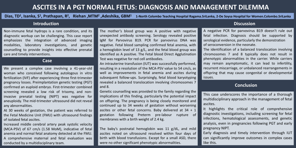
ASCITES IN A PRE IMPLANTATION GENETIC TESTING NORMAL FETUS: DIAGNOSIS AND MANAGEMENT DILEMMA
Dias, TD1, Isanka, S1, Prathapan, R2, Riehan ,MTM2 ,Adeshika, GBM1 1-North Colombo Teaching Hospital Ragama.SriLanka, 2-De Soysa Hospital for Women.Colombo.SriLanka
Introduction
Non-immune fetal hydrops is a rare condition, and its diagnostic workup can be challenging. This case report emphasizes the integration of advanced imaging modalities, laboratory investigations, and genetic counselling to provide insights into effective prenatal care and timely interventions.
Case
We present a complex case involving a 41-year-old woman who conceived following autologous in vitro fertilization (IVF) after experiencing three first-trimester pregnancy losses. Preimplantation genetic testing (PGT) confirmed an euploid embryo. First-trimester combined screening revealed a low risk of trisomy, and non-invasive prenatal testing (NIPT) was negative for aneuploidy. The mid-trimester ultrasound did not reveal any abnormalities.
At 30 weeks of gestation, the patient was referred to the Fetal Medicine Unit (FMU) with ultrasound findings of isolated fetal ascites.
Increased middle cerebral artery peak systolic velocity (MCA-PSV) of 67 cm/s (1.58 MoM), indicative of fetal anemia and normal fetal anatomy detected at the FMU. A comprehensive maternal and fetal evaluation was conducted by a multidisciplinary team.
The mother’s blood group was A positive with negative unexpected antibody screening. Serology revealed positive IgM for Parvovirus B19. PCR for parvovirus DNA was negative. Fetal blood sampling confirmed fetal anemia, with a hemoglobin level of 13 g/L, and the fetal blood group was identified as A positive. The Fetal blood Direct Agglutination Test was negative for red cell antibodies.
An intrauterine transfusion (IUT) was successfully performed, resulting in a resolution of the MCA-PSV value to 54 cm/s, as well as improvements in fetal anemia and ascites during subsequent follow-ups. Surprisingly, fetal blood karyotyping revealed a balanced translocation involving chromosomes 7 and 8.
Genetic counselling was provided to the family regarding the implications of this finding, particularly the potential impact on offspring. The pregnancy is being closely monitored and continued up to 34 weeks of gestation without worsening ascites or other fetal concerns. Baby delivered at 34 + 1 gestation following Preterm pre-labour rupture of membranes with a birth weight of 2.4 kg.
The baby’s postnatal hemoglobin was 11 g/dL, and mild ascites noted on ultrasound resolved within four days of birth with blood transfusions. Except for a small ASD, there were no other significant phenotypic abnormalities.
Discussion
A negative PCR for parvovirus B19 doesn't rule out fetal infection. Diagnosis should be supported by serological evidence, particularly the demonstration
of seroconversion in the neonate.
The identification of a balanced translocation involving chromosomes 7 and 8 typically does not result in phenotypic abnormalities in the carrier. While carriers may remain asymptomatic, it can lead to infertility, recurrent miscarriage, or unbalanced rearrangements in offspring that may cause congenital or developmental issues
Conclusion
This case underscores the importance of a thorough multidisciplinary approach in the management of fetal ascites.
It highlights the critical role of comprehensive diagnostic investigations, including screening for fetal infections, hematological assessments, and genetic analysis, even in pregnancies following PGT and early pregnancy NIPT.
Early diagnosis and timely intervention through IUT can significantly improve outcomes in complex cases like this.