2,281 posters, 63 videos, 56 audios, 43 topics, 18 sessions, 6,174 authors, 2,323 institutions
ePostersLive by SciGen Technologies S.A. All rights reserved.
RCOG World Congress 2025
June 23 - 25, 2025 | ExCeL London, UK
PP.0018
Comparison of ultrasound and magnetic resonance imaging findings in fetal brain anomalies
Poster Presenter
Part of Topic
Fetal medicine
COMPARISON OF ULTRASOUND AND AMGNETIC RESONANCE IMAGING FINDINGS IN THE EVALUATION OF FETAL BRAIN ANOMALIES: A SINGLE INSTITUATION RETROSPECTIVE OBSERVATIONAL STUDY
DR OGULJEMAL REDJEPOVA, ST7 FETAL MEDICINE ATSM
MS PALLAVI KARKHANIS, FETAL MEDICINE CONSULTANT
INTRODUCTION
The International Society of Ultrasound in Obstetrics and Gynaecology provide guidance on the use of fetal MRI to complement ultrasound findings. The multicentre MERIDIAN study found that MRI influenced management of suspected brain abnormalities in more than one third of patients. With improving neurosonography techniques, we evaluated the different conditions in which MRI was used in our unit, focusing on where MRI provided additional information which influenced clinical management.
The aim of this study is to evaluate the role of fetal magnetic resonance imaging (MRI) as a diagnostic adjunct in the management of suspected fetal brain anomalies.
METHODS
All patients who underwent fetal brain MRI between January 2020 and December 2024 in our unit were included. Data was collected from electronic medical records retrospectively.
RESULTS
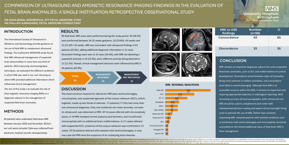
96 fetal brain MRI scans were performed during the study period. 56 (58.3%) were performed between 24-32 weeks gestation, 19 (19.8%) <24 weeks and 21 (21.9)% >32 weeks. MRI was concordant with ultrasound findings in 63 patients (65.6%), adding additional diagnostic information in 11 cases. Discordant findings were seen in 33 cases (34.4%), with MRI not detecting a suspected anomaly in 22 (22.9%), and a different anomaly being detected in 11 (11.5%). Overall, clinical management decisions were influenced by MRI in 44 patients (45.8%).
DISCUSSION
The most common reasons for referral for MRI were ventriculomegaly, microcephaly, and suspected agenesis of the corpus callosum (ACC), which, together, made up two thirds of referrals. 17 patients (17.5%) had more than one ultrasound diagnosis. Only one incidental non-brain anomaly, not seen on ultrasound, was detected on MRI. Of 14 cases referred with microcephaly alone, in 10 MRI revealed normal anatomy and biometry, and 4 confirmed microcephaly with no additional brain malformations. In 21 cases referred with suspected ACC, presence of the corpus callosum was confirmed in 12 cases. Of 24 patients referred with isolated mild ventriculomegaly, in only one case did MRI raise the suspicion of an underlying brain disorder.
CONCLUSION
MRI remains an important diagnostic adjunct for some suspected fetal brain anomalies, such as ACC and malformations of cortical development. Discordance varied between types of anomalies, being most common in midline anomalies, and concordance most likely in ventriculomegaly. Although fetalMRI is an accessible resource within the NHS, it remains an expensive tool, requiring appropriate expertise in radiological reporting. With increasing accuracy of neurosonography, both ultrasound and MRI should be used to complement each other with individualised decision making and expert clinical oversight being used to optimise the use of MRI. Rather than routinely requesting MRI, selected patients with isolated conditions such as borderline-mild ventriculomegaly and microcephaly should be counselled on the limited additional value of fetal brain MRI in their management
1. International Society of Ultrasound in Obstetrics and Gynaecology. ISUOG Practice Guidelines (updated): performance of fetal magnetic resonance imaging. Ultrasound Obstet Gynecol 2023; 61: 278-87. Url: https://www.isuog.org/static/60706d51-b90e-4f9b-80a9008220319e68/Updated-ISUOG-Practice-Guidelines-performance-of-fetal-magnetic-resonance-1.pdf 2. Griffiths P et al. Use fo MRI in the diagnosis of fetal brain abnormalities in utero (MERIDIAN): a multicentre, prospective cohort study. Lancet 2017; 389: 538-46. Url: https://www.thelancet.com/pdfs/journals/lancet/PIIS0140-6736(16)31723-8.pdf 3. Rossi A, Prefumo F. Additional value of fetal magnetic resonance imaging in the prenatal diagnosis of central nervous system anomalies: a systematic review of the literature. Ultrasound Obstet Gynecol 2014; 44(4): 388-93. Url: https://obgyn.onlinelibrary.wiley.com/doi/full/10.1002/uog.13429 4. Hart A et al. Accuracy of in-utero MRI to detect fetal brain abnormalities and prognosticate developmental outcome: postnatal follow-up of the MERIDIAN cohort. Lancet Child Adolesc Health 2020; 4(2): 131-40. Url:https://www.ncbi.nlm.nih.gov/pmc/articles/PMC6988445/ 5. ENSO Working Group. Role of prenatal magnetic resonance imaging in fetuses with isolated mild or moderate ventriculomegaly in the era of neurosonography: international multicenter study. Ultrasound Obstet Gynecol 2020; 56(3): 340-7. Url: https://obgyn.onlinelibrary.wiley.com/doi/10.1002/uog.21974