2,281 posters, 63 videos, 56 audios, 43 topics, 18 sessions, 6,174 authors, 2,323 institutions
ePostersLive by SciGen Technologies S.A. All rights reserved.
RCOG World Congress 2025
June 23 - 25, 2025 | ExCeL London, UK
PP.0012
Novel dermal substitute for skin closure through ≤12Fr fetoscopic cannulas
Poster Presenter
Authors
Jan Deprest, Phililppe De Vloo, Roland Devlieger, Michael Belfort, Luc Joyeux, Hannes van der Merwe, Bart Depreitere, Sarah Devroe, George Attilakos, Sofia Mastrodima-Polychronou, Emma Van den Eede, Paolo De Coppi
Affiliations
Department of Obstetrics and Gynaecology, University Hospitals KU Leuven, Department of Neurosurgery, University Hospitals KU Leuven, Texas Children's Hospital, Department of Anesthesiology, University Hospitals KU Leuven, Institute of Women's Health, University College London
Part of Topic
Fetal medicine
Skin closure of spina bifida using dermal substitutes: need for alternative synthetic dermal patch for repair through 12 Fr fetoscopic cannulas
Jan Deprest1,2, Philippe De Vloo3, Roland Devlieger1, Michael Belfort4, Luc Joyeux4, Hannes Van der Merwe1, Bart Depreitere3, Sara Devroe5, George Attilakos2, Sofia Mastrodima-Polychroniou2, Emma Van den Eede1, Paolo De Coppi1,6,7
1 Department of Obstetrics and Gynaecology, University Hospitals KU Leuven, Leuven, Belgium
2 Institute of Women’s Health, University College London, London, UK
3 Department of Neurosurgery, University Hospitals KU Leuven, Leuven, Belgium
4 Texas Children’s Hospital, Houston, Texas
5 Department of Anesthesiology, University Hospitals Leuven, Leuven, Belgium
6 Institute of Child Health, University College London, London, U.K.
7 Great Ormond Street Hospital for Children, London, U.K.
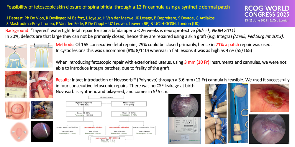
Objective: In selected cases, fetal spina bifida repair improves outcome as compared to postnatal repair. The surgery requires creation of a multi-layered watertight repair, which may be challenging in large lesions. There is an increasing demand for fetoscopic repair, even though this remains investigational. In our program we aim to mimic the open procedure as much as possible. In case of large skin defects, we use a dermal substitute rather than relaxing incisions or plastic surgery techniques. Because xenogenic dermal collagen matrix (Integra) becomes damaged by insertion through the largest 12 Fr (3.6 mm) fetoscopic cannula used, we moved to the synthetic NovoSorb patch, which is more robust.
Design: Prospective observational study on 165 consecutive fetal spina bifida repairs in patients meeting the criteria of the “Management Of Myelomeningocele Study”. The goal was to determine the relation between lesion type and patch use and to report on the feasibility of patch use through a 12 Fr cannula.
Methods: This is a retrospective analysis of prospectively collected data on 165 consecutive fetal spina bifida repairs, including 22 consecutive fetoscopic and 143 repairs by (mini-)hysterotomy, all under general fetomaternal anesthesia. Outcomes were feasibility of intact insertion and suturing and postoperative complications till the end of the neonatal period.
Results: 67% (110/165) of lesions were cystic, leaving 55 myeloschisis cases (33%). The overall skin patch rate was 34/165 (21%), including four out of 22 operated by fetoscopy (17%; NS). Skin grafts were more frequently required in myeloschisis (47%) compared to myelomeningocele (7%; p=1*10-9). Intact introduction of Integra dermal patch was only possible following cannula removal and enlargement of the entry point. Conversely, we successfully introduced a Novosorb-patch (n=3) without compromising its structure. The patch was successfully sutured in place. There was no leakage at birth and no local complications were reported (n=20 born).
Conclusion: NovoSorb® is a synthetic skin substitute that resists damage during insertion through a 12 Fr cannula and has good suture retention capability. This allows us to offer the same neurosurgical technique to patients who choose fetoscopy. No short term local complications to his patch were observed. Long term studies should determine the risk for epidermoid inclusion cysts and wound dehiscence.