2,281 posters, 63 videos, 56 audios, 43 topics, 18 sessions, 6,174 authors, 2,323 institutions
ePostersLive by SciGen Technologies S.A. All rights reserved.
RCOG World Congress 2025
June 23 - 25, 2025 | ExCeL London, UK
EP.2179
Uncommon fibroids:A diagnostic challange
Poster Presenter
Authors
Affiliations
Part of Topic
Urogynaecology/pelvic floor
Introduction
Leiomyomas are benign fibromuscular tumors of the smooth muscle cells, thus mesenchymal in origin and found to be estrogen dependant. It broadly divided into subserosal, intramural, and submucosal types. Uncommon locations are in the cervix, uterosacral ligaments, round ligament, ovaries, and inguinal canal. Vaginal leiomyoma is said to be the rarest of all. Approximately, the vaginal paraurethral leiomyomas represent 5 % of all paraurethral masses, which 1:1000 women.1 It is seen mostly among women in the age group of 35 to 50 years, mostly among Caucasians. The diagnosis and management itself become challenging due to its uncommon location, and its anatomic proximity to urethra, bladder and vagina. Here we have discussed 2 such cases of diagnostic challenge, which we have identified and successfully managed by surgical excision of a vaginal and a paraurethral fibroid.
CASE 1:
A 37-year-old female, P1L1, visited the gynecology outpatient department with the chief complaints of an irreducible mass protruding from the vaginal introitus. She had difficulty in micturation and often had incomplete evacuation of the bladder and urinary retention.
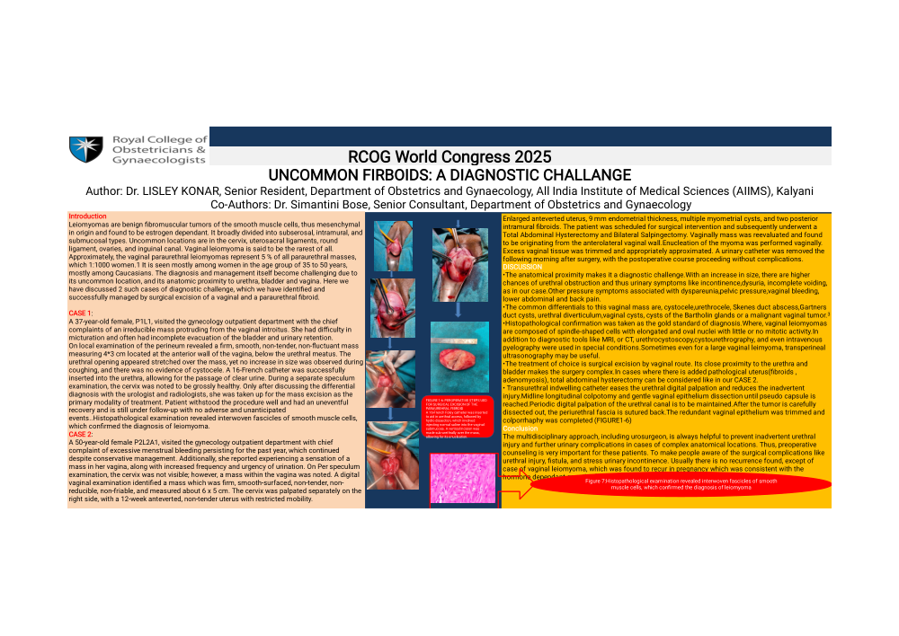
On local examination of the perineum revealed a firm, smooth, non-tender, non-fluctuant mass measuring 4*3 cm located at the anterior wall of the vagina, below the urethral meatus. The urethral opening appeared stretched over the mass, yet no increase in size was observed during coughing, and there was no evidence of cystocele. A 16-French catheter was successfully inserted into the urethra, allowing for the passage of clear urine. During a separate speculum examination, the cervix was noted to be grossly healthy. Only after discussing the differential diagnosis with the urologist and radiologists, she was taken up for the mass excision as the primary modality of treatment. Patient withstood the procedure well and had an uneventful recovery and is still under follow-up with no adverse and unanticipated events..Histopathological examination revealed interwoven fascicles of smooth muscle cells, which confirmed the diagnosis of leiomyoma.
CASE 2:
A 50-year-old female P2L2A1, visited the gynecology outpatient department with chief complaint of excessive menstrual bleeding persisting for the past year, which continued despite conservative management. Additionally, she reported experiencing a sensation of a mass in her vagina, along with increased frequency and urgency of urination. On Per speculum examination, the cervix was not visible; however, a mass within the vagina was noted. A digital vaginal examination identified a mass which was firm, smooth-surfaced, non-tender, non-reducible, non-friable, and measured about 6 x 5 cm. The cervix was palpated separately on the right side, with a 12-week anteverted, non-tender uterus with restricted mobility.Enlarged anteverted uterus, 9 mm endometrial thickness, multiple myometrial cysts, and two posterior intramural fibroids. The patient was scheduled for surgical intervention and subsequently underwent a Total Abdominal Hysterectomy and Bilateral Salpingectomy. Vaginally mass was reevaluated and found to be originating from the anterolateral vaginal wall.Enucleation of the myoma was performed vaginally. Excess vaginal tissue was trimmed and appropriately approximated. A urinary catheter was removed the following morning after surgery, with the postoperative course proceeding without complications.
DISCUSSION
The anatomical proximity makes it a diagnostic challenge.With an increase in size, there are higher chances of urethral obstruction and thus urinary symptoms like incontinence,dysuria, incomplete voiding,as in our case.Other pressure symptoms associated with dyspareunia,pelvic pressure,vaginal bleeding, lower abdominal and back pain.
The common differentials to this vaginal mass are, cystocele,urethrocele, Skenes duct abscess,Gartners duct cysts, urethral diverticulum,vaginal cysts, cysts of the Bartholin glands or a malignant vaginal tumor.3
Histopathological confirmation was taken as the gold standard of diagnosis.Where, vaginal leiomyomas are composed of spindle-shaped cells with elongated and oval nuclei with little or no mitotic activity.In addition to diagnostic tools like MRI, or CT, urethrocystoscopy,cystourethrography, and even intravenous pyelography were used in special conditions.Sometimes even for a large vaginal leimyoma, transperineal ultrasonography may be useful.
The treatment of choice is surgical excision by vaginal route. Its close proximity to the urethra and bladder makes the surgery complex.In cases where there is added pathological uterus(fibroids , adenomyosis), total abdominal hysterectomy can be considered like in our CASE 2.
Transurethral indwelling catheter eases the urethral digital palpation and reduces the inadvertent injury.Midline longitudinal colpotomy and gentle vaginal epithelium dissection until pseudo capsule is reached.Periodic digital palpation of the urethral canal is to be maintained.After the tumor is carefully dissected out, the periurethral fascia is sutured back.The redundant vaginal epithelium was trimmed and colporrhaphy was completed (FIGURE1-6)
Conclusion
The multidisciplinary approach, including urosurgeon, is always helpful to prevent inadvertent urethral injury and further urinary complications in cases of complex anatomical locations. Thus, preoperative counseling is very important for these patients. To make people aware of the surgical complications like urethral injury, fistula, and stress urinary incontinence. Usually there is no recurrence found, except of case of vaginal leiomyoma, which was found to recur in pregnancy which was consistent with the hormone dependant growth of the tumor.