2,281 posters, 63 videos, 56 audios, 43 topics, 18 sessions, 6,174 authors, 2,323 institutions
ePostersLive by SciGen Technologies S.A. All rights reserved.
RCOG World Congress 2025
June 23 - 25, 2025 | ExCeL London, UK
EP.1852
Audit on the Management of Ovarian Torsion Cases at Ipswich Hospital
Poster Presenter
Authors
Affiliations
Part of Topic
Quality improvement
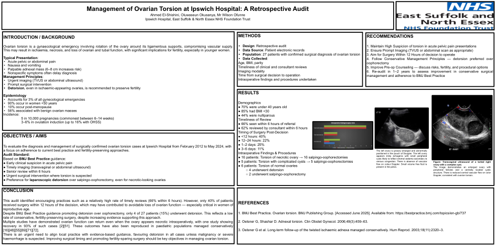
Management of Ovarian Torsion at Ipswich Hospital: A Retrospective Audit Ahmed El-Shishini, Oluwaseun Okusanya, Mr Wilson Ofunne Ipswich Hospital, East Suffolk & North Essex NHS Foundation Trust METHODS • Design: Retrospective audit • Data Source: Patient electronic records • Population: 27 patients with confirmed surgical diagnosis of ovarian torsion • Data Collected: Age, BMI, parity Timeliness of clinical and consultant reviews Imaging modality Time from surgical decision to operation Intraoperative findings and procedures undertaken OBJECTIVES / AIMS To evaluate the diagnosis and management of surgically confirmed ovarian torsion cases at Ipswich Hospital from February 2012 to May 2024, with a focus on adherence to current best practice and fertility-preserving approaches. Audit Standard: Based on BMJ Best Practice guidance: ● Early clinical suspicion in acute pelvic pain ● Timely imaging (transvaginal or abdominal ultrasound) ● Senior review within 6 hours ● Urgent surgical intervention where torsion is suspected ● Preference for laparoscopic detorsion over salpingo-oophorectomy, even for necrotic-looking ovaries INTRODUCTION / BACKGROUND Ovarian torsion is a gynaecological emergency involving rotation of the ovary around its ligamentous supports, compromising vascular supply. This may result in ischaemia, necrosis, and loss of ovarian and tubal function, with significant implications for fertility, especially in younger women. Typical Presentation: • Acute pelvic or abdominal pain • Nausea and vomiting • Palpable adnexal mass (6–8 cm increases risk) • Nonspecific symptoms often delay diagnosis Management Principles: • Urgent imaging (TVUS or abdominal ultrasound) • Prompt surgical intervention • Detorsion, even in ischaemic-appearing ovaries, is recommended to preserve fertility Epidemiology • Accounts for 3% of all gynecological emergencies • 90% occur in women <50 years • 10% occur post-menopause • 56% associated with benign ovarian masses Incidence: 5 in 10,000 pregnancies (commonest between 6–14 weeks) 3–6% in ovulation induction (up to 16% with OHSS) CONCLUSION This audit identified encouraging practices such as a relatively high rate of timely reviews (66% within 6 hours). However, only 40% of patients received surgery within 12 hours of the decision, which may have contributed to avoidable loss of ovarian function — especially critical in women of reproductive age. Despite BMJ Best Practice guidance promoting detorsion over oophorectomy, only 4 of 27 patients (15%) underwent detorsion. This reflects a low rate of conservative, fertility-preserving surgery, despite increasing evidence supporting this approach. Multiple studies have demonstrated ovarian function can return even when the ovary appears necrotic intraoperatively, with one study showing recovery in 93% of such cases [2][51]. These outcomes have also been reproduced in paediatric populations managed conservatively [16][48][55][69][71][72]. There is an urgent need to align local practice with evidence-based guidance, favouring detorsion in all cases unless malignancy or severe haemorrhage is suspected. Improving surgical timing and promoting fertility-sparing surgery should be key objectives in managing ovarian torsion. RESULTS Demographics ● 70% were under 40 years old ● 85% had BMI <30 ● 44% were nulliparous Timeliness of Review ● 66% seen within 6 hours of referral ● 62% reviewed by consultant within 6 hours Timing of Surgery Post-Decision ● <12 hours: 40% ● 12–24 hours: 22% ● 1–2 days: 25% ● 3–6 days: 11% Intraoperative Findings & Procedures ● 16 patients: Torsion of necrotic ovary → 16 salpingo-oophorectomies ● 5 patients: Torsion with complicated cysts → 5 salpingo-oophorectomies ● 6 patients: Torsion of normal ovaries ○ 4 underwent detorsion ○ 2 underwent salpingo-oophorectomy The left ovary is grossly enlarged and abnormally positioned in the pouch of Douglas. The left ovary appears mildy echogenic with small peripheral cysts likely to reflect stromal oedema secondary to venous congestion. There is absence of vascular flow on colour Doppler. Small volume free fluid is present in the pelvis. Figure: Transvaginal ultrasound of a torted right ovary with a complex cyst. The image demonstrates an enlarged ovary with peripheral follicles and a centrally located cystic structure. There is reduced central vascular flow on color Doppler, consistent with ovarian torsion. RECOMMENDATIONS 1. Maintain High Suspicion of torsion in acute pelvic pain presentations 2. Ensure Prompt Imaging (TVUS or abdominal scan as appropriate) 3. Aim for Surgery Within 12 Hours of decision to operate 4. Follow Conservative Management Principles — detorsion preferred over oophorectomy 5. Improve Pre-op Counseling — discuss risks, fertility, and procedural options 6. Re-audit in 1–2 years to assess improvement in conservative surgical management and adherence to BMJ Best Practice REFERENCES 1. BMJ Best Practice. Ovarian torsion. BMJ Publishing Group. [Accessed June 2025]. Available from: https://bestpractice.bmj.com/topics/en-gb/737 2. Oelsner G, Shashar D. Adnexal torsion. Clin Obstet Gynecol. 2006;49(3):459–63. 3. Oelsner G et al. Long-term follow-up of the twisted ischaemic adnexa managed conservatively. Hum Reprod. 2003;18(11):2320–3