2,281 posters, 63 videos, 56 audios, 43 topics, 18 sessions, 6,174 authors, 2,323 institutions
ePostersLive by SciGen Technologies S.A. All rights reserved.
RCOG World Congress 2025
June 23 - 25, 2025 | ExCeL London, UK
EP.1807
Pure Sertoli Cell Ovarian Tumour: A rare cause of post-menopausal adnexal torsion
Poster Presenter
Authors
Affiliations
Part of Topic
Post reproductive health
INTRODUCTION
We present a report with multiple rarities in a postmenopausal woman. Adnexal torsion is uncommon in postmenopausal women (PMW), with the finding of malignancy being <3% [1]. Sertoli-Leydig cell tumours (SLCTs) are extremely rare,and less than 10% present post-menopause. SLCTs are typically unilateral and are confined to the ovary at the time of diagnosis [2]. Pure Sertoli cell tumours (PSCTs) are rarer still, accounting for <4% SLCTs [2].
CASE HISTORY
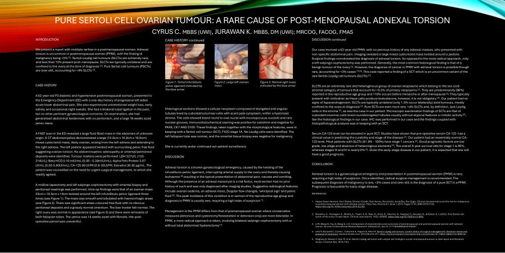
A 62-year-old P3 diabetic and hypertensive postmenopausal woman, presented to the Emergency Department (ED) with a one-day history of progressive left sided acute lower abdominal pain. She also experienced unintentional weight loss, early satiety and occasional night sweats. She had a bilateral tubal ligation in the past, but no other pertinent gynaecological concerns. On examination, she had generalised abdominal tenderness with no peritonism, and a large 16 weeks sized pelvic mass. A FAST scan in the ED revealed a large fluid filled mass in the abdomen of unknown origin. A CT abdomen/pelvis demonstrated a large (13.0cm x 16.9cm x 18.9cm) mixed cystic/solid mass, likely ovarian, arising from the left adnexa and extending to the right adnexa. The left pedicle appeared twisted with surrounding pelvic free fluid suggesting ovarian torsion. No abdominopelvic adenopathy or omental/peritoneal deposits were identified. Tumour markers were performed: LDH 327U/L (135-214U/L); Beta HCG 0.10 mIU/mL (0.00 – 5.30mIU/mL); Alpha Feto Protein 3.87 UI/mL (0.50-5.80UI/mL); CA-125 36 UI/MI (0.6-35UI/MI; Estradiol 30.92 pg/mL. The patient was counselled on the need for urgent surgical management, to which she readily agreed. A midline laparotomy and left salpingo-oophorectomy with omental biopsy and peritoneal washings was performed. Intra-op findings were that of an ovarian mass 23cm x 18.5cm x 14cm twisted around the left infundibulo-pelvic ligament three times (see Figure 1). The mass was smooth and lobulated with haemorrhagic areas (see Figure 2). There was significant straw-coloured free fluid with no obvious peritoneal deposits and a grossly normal omentum. The liver border felt normal. The right ovary was normal in appearance (see Figure 3) and there were remnants of both fallopian tubes. The uterus was 14 weeks sized with fibroids. Her post-operative period was uneventful.
Figure 1: Torted infundibulo- pelvic ligament indicated by the blue arrow
Figure 2: Large left ovarian mass
Figure 3: Normal right ovary
Our case involved a 62-year-old PMW, with no previous history of any adnexal masses, who presented with non-specific abdominal pain. Imaging revealed a large mixed cystic/solid mass twisted around a pedicle. Surgical findings corroborated the diagnosis of adnexal torsion. As opposed to the more radical approach, only a left salpingo-oophorectomy was performed. Generally, the most common histological finding is that of a benign tumour of the ovary [3]. However, the diagnosis of cancer in PMW with adnexal torsion is possible though rare, accounting for <3% cases [1] [4]. This case reported a finding of a SCT which is an uncommon variant of the rare Sertoli-Leydig cell tumours (SLCTs) [2].
SLCTS are an extremely rare and heterogenous group of ovarian neoplasms which belong to the sex cord-stromal category of tumours that account for <0.5% of primary neoplasms [2]. They are predominantly (90%) reported in the reproductive age group and <10% occurs before menarche or after menopause [2]. They typically present with virilization as these cells produce testosterone, however, it is not obligatory [2]. Our patient had no signs of hyperandrogenism. SLCTs are typically unilateral (only 1.5% occur bilaterally) solid tumours, mostly confined to the ovary at diagnosis [2]. Pure SCTs are even more rare, <4% SLCTs and, by definition, lack Leydig cells in the stroma [2], as was the case in our patient. Microscopic examination findings of SCTs are that of cuboidal/columnar cells lined round/elongated tubules usually without atypical features or mitotic activity [2], like the histological findings in our case. IHC was performed in our case and the findings coupled with histopathological analysis was in keeping with an SCT. Histological sections showed a cellular neoplasm composed of elongated and angular tubules lined by cuboidal/columnar cells with scant pale cytoplasm, within a hyalinized stroma. The cells showed bland round to oval nuclei with inconspicuous nucleoli and rare nuclear grooves. Immunohistochemical staining was positive for calretinin and negative for PAX8, CK7 AND S100. These findings, taken together with the morphological features, were in keeping with a Sertoli cell tumour (SCT), FIGO stage 1A. No Leydig cells were identified. The left fallopian tube was normal, and the omental tissue biopsy was negative for malignancy.
She is currently under continued out-patient surveillance.
DISCUSSION
Adnexal torsion is a known gynaecological emergency, caused by the twisting of the infundibulo-pelvic ligament, interrupting arterial supply to the ovary and thereby causing ischaemia [2] resulting in the typical presentation of abdominal pain, nausea and vomiting. Although the presence of an adnexal mass/cyst is a risk factor, most women had no prior history of such and was only diagnosed after imaging studies. Suggestive radiological features include ovarian oedema, an adnexal mass, Doppler flow changes, ‘whirlpool sign’ and pelvic fluid [3]. The peak incidence of this condition is in women of the reproductive age group and diagnosis in PMW is usually rare, requiring a high index of suspicion [1]. Management in the PMW differs from that of premenopausal women where conservative measures (detorsion and cystectomy/fenestration or detorsion only) are more desirable. In PMW, a more radical approach is taken, involving bilateral salpingo-oophorectomy with or without total abdominal hysterectomy [1]. Serum CA-125 level can be elevated in pure SCT. Studies have shown that pre-operative serum CA-125 has a clinical value in predicting the curability and stage of the disease [2]. Our patient had an essentially normal CA-125 level. Most patients with SLCTs (81.8% - 100%) have stage 1 cancers [2]. Good prognostic factors are low grade, low stage and absence of heterologous elements [2]. The overall 5-year survival rate for stage 1 is 95%, whereas stages III and IV is nearly 0% [5]. Given the early-stage disease in our patient, it is expected that she will have a good prognosis.
CONCLUSION
Adnexal torsion is a gynaecological emergency and presentation in postmenopausal women (PMW) is rare, requiring a high index of suspicion. Once identified, radical surgical management is recommended. The subsequent diagnosis of malignancy is rare, <3% cases and rarer still is the diagnosis of a pure SCT in a PMW.
Prognosis is favourable for early-stage disease.
REFERENCES
1. Hadas Ganer Herman, Amir Shalev, Shimon Ginath, Ram Kerner, Ran Keidar, Jacob Bar, Ron Sagiv, Clinical characteristics and the risk for malignancy in postmenopausal women with adnexal torsion, Maturitas, Volume 81, Issue 1,2015, Pages 57-61,ISSN 0378-5122, https://doi.org/10.1016/j.maturitas.2015.02.261.
2. Shrestha, S., Homagain, S., Bhatta, S., Tiwari, S. B., Rijal, R., Aryal, R., Sharma, N., Paudyal, P., Katuwal, N., & Rawal, S. J. (2022). Pure Sertoli cell tumor of the ovary: A case report. Clinical case reports, 10(5), e05892. https://doi.org/10.1002/ccr3.5892
3. Li M, Wang H, Hou S, Wang S, Li H. Comparison of characteristics and outcomes of premenopausal and postmenopausal women with adnexaltorsion. Journal of International Medical Research. 2024;52(12). doi:10.1177/03000605241305251
4. Litta P, Saccardi C, Conte L, Codroma A, Angioni S, Mioni R. Sertoli-Leydig cell tumors: current status of surgical management: literature review andproposal of treatment. Gynecol Endocrinol. 2013;29:412–417. https://ovarianresearch.biomedcentral.com/articles/10.1186/s13048-014-0083-52.
5. Moghazy D, Sharan C, Nair M, et al. Sertoli-Leydig cell tumor with unique nail findings in a post-menopausal woman: a case report and literaturereview. J Ovarian Res. 2014;7:83.