2,281 posters, 63 videos, 56 audios, 43 topics, 18 sessions, 6,174 authors, 2,323 institutions
ePostersLive by SciGen Technologies S.A. All rights reserved.
RCOG World Congress 2025
June 23 - 25, 2025 | ExCeL London, UK
EP.1789
When Gynaecology and Paediatrics collaborate: Inguinal Herniation of Ovary in a Neonate.
Poster Presenter
Authors
Affiliations
Part of Topic
Paediatric & adolescent gynaecology
When Gynaecology and Paediatrics collaborate: Inguinal Herniation of Ovary in a Neonate.
By Sara Kidher¹, Dr Hayat Alraeesi², Dr Saima Qayoom²
INTRODUCTION:
This case report will discuss an incident regarding an inguinal hernia, a common epidemiology that affects over 25% of men (Hammoud and Gerken, 2023). Interestingly, the same pathology applied to infant females appears to be much less common. To further explore uncommon incidents that occur with inguinal hernias; this report describes a case of an inguinal hernia containing the adnexa of a 3 week old neonate. With this in mind, a multifaceted clinical picture is painted, where gynaecology and paediatrics, alongside radiology must all play their role.
CASE REPORT:
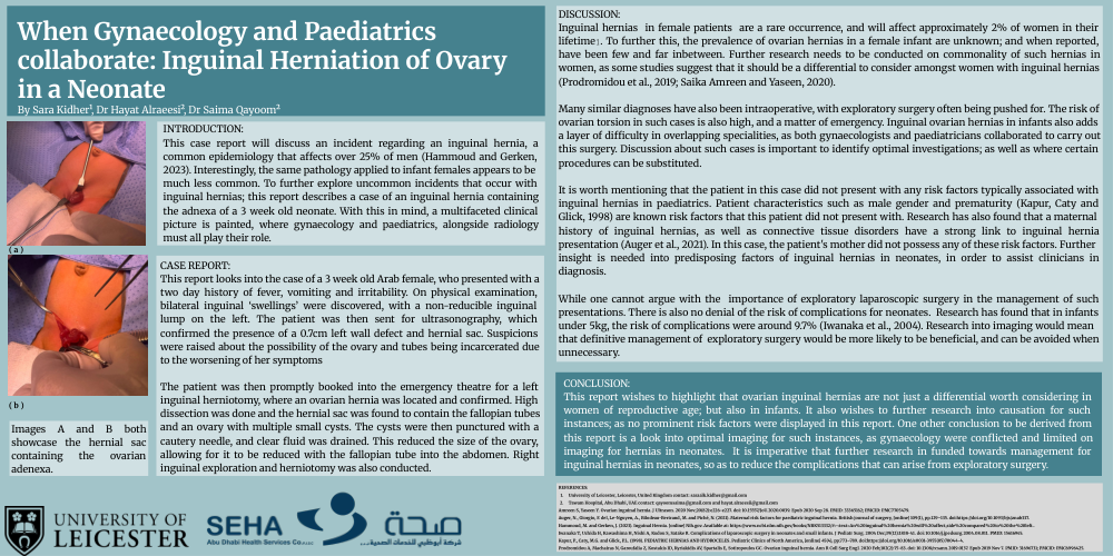
This report looks into the case of a 3 week old Arab female, who presented with a two day history of fever, vomiting and irritability. On physical examination, bilateral inguinal ‘swellings’ were discovered, with a non-reducible inguinal lump on the left. The patient was then sent for ultrasonography, which confirmed the presence of a 0.7cm left wall defect and hernial sac. Suspicions were raised about the possibility of the ovary and tubes being incarcerated due to the worsening of her symptoms
The patient was then promptly booked into the emergency theatre for a left inguinal herniotomy, where an ovarian hernia was located and confirmed. High dissection was done and the hernial sac was found to contain the fallopian tubes and an ovary with multiple small cysts. The cysts were then punctured with a cautery needle, and clear fluid was drained. This reduced the size of the ovary, allowing for it to be reduced with the fallopian tube into the abdomen. Right inguinal exploration and herniotomy was also conducted.
DISCUSSION:
Inguinal hernias in female patients are a rare occurrence, and will affect approximately 2% of women in their lifetime⥠. To further this, the prevalence of ovarian hernias in a female infant are unknown; and when reported, have been few and far inbetween. Further research needs to be conducted on commonality of such hernias in women, as some studies suggest that it should be a differential to consider amongst women with inguinal hernias (Prodromidou et al., 2019; Saika Amreen and Yaseen, 2020).
Many similar diagnoses have also been intraoperative, with exploratory surgery often being pushed for. The risk of ovarian torsion in such cases is also high, and a matter of emergency. Inguinal ovarian hernias in infants also adds a layer of difficulty in overlapping specialities, as both gynaecologists and paediatricians collaborated to carry out this surgery. Discussion about such cases is important to identify optimal investigations; as well as where certain procedures can be substituted.
It is worth mentioning that the patient in this case did not present with any risk factors typically associated with inguinal hernias in paediatrics. Patient characteristics such as male gender and prematurity (Kapur, Caty and Glick, 1998) are known risk factors that this patient did not present with. Research has also found that a maternal history of inguinal hernias, as well as connective tissue disorders have a strong link to inguinal hernia presentation (Auger et al., 2021). In this case, the patient's mother did not possess any of these risk factors. Further insight is needed into predisposing factors of inguinal hernias in neonates, in order to assist clinicians in diagnosis.
While one cannot argue with the importance of exploratory laparoscopic surgery in the management of such presentations. There is also no denial of the risk of complications for neonates. Research has found that in infants under 5kg, the risk of complications were around 9.7% (Iwanaka et al., 2004). Research into imaging would mean that definitive management of exploratory surgery would be more likely to be beneficial, and can be avoided when unnecessary.
CONCLUSION:
This report wishes to highlight that ovarian inguinal hernias are not just a differential worth considering in women of reproductive age; but also in infants. It also wishes to further research into causation for such instances; as no prominent risk factors were displayed in this report. One other conclusion to be derived from this report is a look into optimal imaging for such instances, as gynaecology were conflicted and limited on imaging for hernias in neonates. It is imperative that further research in funded towards management for inguinal hernias in neonates, so as to reduce the complications that can arise from exploratory surgery.
REFERENCES:
1. 2. University of Leicester, Leicester, United Kingdom contact: saraalk.kidher@gmail.com
Tawam Hospital, Abu Dhabi, UAE contact: qayoomsaima@gmail.com and hayat.alraeesii@gmail.com
Amreen S, Yaseen Y. Ovarian inguinal hernia. J Ultrason. 2020 Nov;20(82):e226-e227. doi: 10.15557/JoU.2020.0039. Epub 2020 Sep 28. PMID: 33365162; PMCID: PMC7705479.
Auger, N., Giorgio, F. del, Le-Nguyen, A., Bilodeau-Bertrand, M. and Piché, N. (2021). Maternal risk factors for paediatric inguinal hernia. British journal of surgery, [online] 109(1), pp.129–135. doi:https://doi.org/10.1093/bjs/znab337.
Hammoud, M. and Gerken, J. (2023). Inguinal Hernia. [online] Nih.gov. Available at: https://www.ncbi.nlm.nih.gov/books/NBK513332/#:~:text=An%20inguinal%20hernia%20will%20affect,side%20compared%20to%20the%20left..
Iwanaka T, Uchida H, Kawashima H, Nishi A, Kudou S, Satake R. Complications of laparoscopic surgery in neonates and small infants. J Pediatr Surg. 2004 Dec;39(12):1838-41. doi: 10.1016/j.jpedsurg.2004.08.011. PMID: 15616945.
Kapur, P., Caty, M.G. and Glick, P.L. (1998). PEDIATRIC HERNIAS AND HYDROCELES. Pediatric Clinics of North America, [online] 45(4), pp.773–789. doi:https://doi.org/10.1016/s0031-3955(05)70044-4.
Prodromidou A, Machairas N, Garoufalia Z, Kostakis ID, Kyriakidis AV, Spartalis E, Sotiropoulos GC. Ovarian inguinal hernia. Ann R Coll Surg Engl. 2020 Feb;102(2):75-83. doi: 10.1308/rcsann.2019.0137. Epub 2019 Nov 7. PMID: 31696731; PMCID: PMC6996425.