1,830 posters, 67 videos, 75 audios, 29 topics, 22 sessions, 4,727 authors, 1,697 institutions
ePostersLive by SciGen Technologies S.A. All rights reserved.
RCOG World Congress 2023
June 12 - 14, 2023 | London, UK

EP.0506
Understanding the Peritoneal Micro-environment in Endometriosis
Poster Presenter
Authors
Harriet Lamb, Debbie Fischer, Gaity Ahmad, Kay Marshall, Jessica Traynor, Emma Crosbie
Affiliations
Part of Topic
Endometriosis and Uterine Disorders
Understanding the Peritoneal Micro-environment in Endometriosis
What is the Peritoneum?
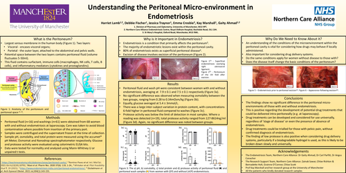
•Largest serous membrane in the human body (Figure 1). Two layers:
•Visceral - encases visceral organs;
•Parietal - the outer layer, attached to the abdominal and pelvic walls.
•Potential space between the two layers contains peritoneal fluid (volume fluctuates 5-50ml).
•This fluid contains surfactant, immune cells (macrophages, NK cells, T cells, B cells), and inflammatory mediators (cytokines and prostaglandins).
Why is it Important in Endometriosis?
•Endometriosis is a condition that primarily affects the peritoneum3.
•The majority of endometriotic lesions exist within the peritoneal cavity.
•80% of endometriosis exists as superficial peritoneal disease4.
Excision of disease involves excision of the peritoneum (Figure 2).
Why Do We Need to Know About it?
•An understanding of the conditions of the microenvironment within the peritoneal cavity is vital for considering how drugs may behave if locally administered.
•Also important for considering drug delivery systems.
•Do the same conditions apply for women without disease to those with?
•Does the disease itself change the basic conditions of the peritoneum?
Methods
•Peritoneal fluid (n=16) and washings (n=61) were obtained from 68 women with and without endometriosis at laparoscopy. Care was taken to avoid blood contamination where possible from insertion of the primary port.
•Samples were centrifuged and the supernatant frozen at the time of collection.
•Sample pH, osmolality, and total protein were measured using the accumet® pH Meter, Osmomat and Nanodrop spectrophotometer respectively. Glucose and protease activity were evaluated using colorimetric ELISA kits.
•Data were tested for normality and analyzed using Mann-Whitney U or unpaired t-tests.
Results
•Peritoneal fluid and wash pH were consistent between women with and without endometriosis, averaging at 7.9 ± 0.1 and 7.5 ± 0.1 respectively (Figure 3a).
•No significant difference was observed when measuring osmolality between the two groups, ranging from 0.28 to 0.34mOsm/kg (Figure 3b).
•Equally, glucose averaged at 5.4 ± 3mmol/L.
•There was a large inter-subject variation in protein content, with concentrations 6-fold higher in peritoneal fluid compared to washes (Figure 3c).
•Protease activity was below the limit of detection in most samples. Where a reading was detected (n=19), total protease activity ranged from 127-8614ng/ml (Figure 3d). Again, no significant difference was noted between groups.
Conclusions
•The findings show no significant difference in the peritoneal micro-environments of those with and without endometriosis.
•This is positive regarding the development of potential drug treatments that could be delivered intra-peritoneally (e.g. at laparoscopy).
•Drug treatments can be developed and considered for use universally, regardless of ‘stage of disease’ or even the presence of absence of endometriosis.
•Drug treatments could be trialled for those with pelvic pain, without confirmed diagnosis of endometriosis.
•The finding of low protease is also positive when considering drug delivery systems, particularly if a biodegradable hydrogel is used, as this is likely to be broken down slowly and universally.