1,830 posters, 67 videos, 75 audios, 29 topics, 22 sessions, 4,727 authors, 1,697 institutions
ePostersLive by SciGen Technologies S.A. All rights reserved.
RCOG World Congress 2023
June 12 - 14, 2023 | London, UK

EP.0389
Diagnosis of Leydig Cell Tumour following presentation with virilisation and ovarian hyperthecosis
Poster Presenter
Part of Topic
Post Reproductive Health
Diagnosis of Leydig Cell Tumour following presentation with virilization and ovarian hyperthecosis
William Wilson-Theaker1 , Nora Ahmed2 , Jagadeeswari Karuppaswamy2, Sushmita Pearce3.
1Specialist Registrar 2Obstetrics and Gynaecology Consultant 3Diabetes and Endocrinology Consultant
WWL Teaching Hospitals NHS Foundation Trust
Background
Leydig cell ovarian tumour is a rare sex cord-gonadal stromal tumour which constitutes less than 0.5% of ovarian tumours1. These cancers appear at all ages but most of the cases concern women in the perimenopause. Leydig cell tumours are difficult to diagnose as they are often difficult to detect despite multiple different imaging studies2.
Case
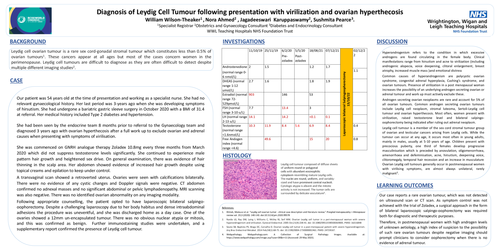
Our patient was 54 years old at the time of presentation and working as a specialist nurse. She had no relevant gynaecological history. Her last period was 3-years ago when she was developing symptoms of hirsutism. She had undergone a bariatric gastric sleeve surgery in October 2020 with a BMI of 31.4 at referral. Her medical history included Type 2 diabetes and hypertension.
She had been seen by the endocrine team 8 months prior to referral to the Gynaecology team and diagnosed 3 years ago with ovarian hyperthecosis after a full work up to exclude ovarian and adrenal causes when presenting with symptoms of virilisation.
She was commenced on GNRH analogue therapy Zoladex 10.8mg every three months from March 2020 which did not suppress testosterone levels significantly. She continued to experience male pattern hair growth and heightened sex drive. On general examination, there was evidence of hair thinning in the scalp area. Her abdomen showed evidence of increased hair growth despite using topical creams and epilation to keep under control.
A transvaginal scan showed a retroverted uterus. Ovaries were seen with calcifications bilaterally. There were no evidence of any cystic changes and Doppler signals were negative. CT abdomen confirmed no adnexal masses and no significant abdominal or pelvic lymphadenopathy. MRI scanning was also negative. There was no identified ovarian abnormality on any imaging modality.
Following appropriate counselling, the patient opted to have laparoscopic bilateral salpingo-oophorectomy. Despite a challenging laparoscopy due to her body habitus and dense intraabdominal adhesions the procedure was uneventful, and she was discharged home as a day case. One of the ovaries showed a 12mm un-encapsulated tumour. There was no obvious nuclear atypia or mitosis, and this was confirmed as benign. Further immunostaining studies were undertaken, and a supplementary report confirmed the presence of Leydig cell tumour.
Discussion
Our patient was 54 years old at the time of presentation and working as a specialist nurse. She had no relevant gynaecological history. Her last period was 3-years ago when she was developing symptoms of hirsutism. She had undergone a bariatric gastric sleeve surgery in October 2020 with a BMI of 31.4 at referral. Her medical history included Type 2 diabetes and hypertension.
She had been seen by the endocrine team 8 months prior to referral to the Gynaecology team and diagnosed 3 years ago with ovarian hyperthecosis after a full work up to exclude ovarian and adrenal causes when presenting with symptoms of virilisation.
She was commenced on GNRH analogue therapy Zoladex 10.8mg every three months from March 2020 which did not suppress testosterone levels significantly. She continued to experience male pattern hair growth and heightened sex drive. On general examination, there was evidence of hair thinning in the scalp area. Her abdomen showed evidence of increased hair growth despite using topical creams and epilation to keep under control.
A transvaginal scan showed a retroverted uterus. Ovaries were seen with calcifications bilaterally. There were no evidence of any cystic changes and Doppler signals were negative. CT abdomen confirmed no adnexal masses and no significant abdominal or pelvic lymphadenopathy. MRI scanning was also negative. There was no identified ovarian abnormality on any imaging modality.
Following appropriate counselling, the patient opted to have laparoscopic bilateral salpingo-oophorectomy. Despite a challenging laparoscopy due to her body habitus and dense intraabdominal adhesions the procedure was uneventful, and she was discharged home as a day case. One of the ovaries showed a 12mm un-encapsulated tumour. There was no obvious nuclear atypia or mitosis, and this was confirmed as benign. Further immunostaining studies were undertaken, and a supplementary report confirmed the presence of Leydig cell tumour.
Learning Outcomes
Our case reports a rare ovarian tumour, which was not detected on ultrasound scan or CT scan. As symptom control was not achieved with the trial of Zoladex, a surgical approach in the form of bilateral laparoscopic salpingo-oophorectomy was required both for diagnostic and therapeutic purposes.
Therefore, in postmenopausal women with high androgen levels of unknown aetiology, a high index of suspicion to the possibility of such rare ovarian tumours despite negative imaging should prompt clinicians to consider oophorectomy when there is no evidence of adrenal tumour.
References: