1,830 posters, 67 videos, 75 audios, 29 topics, 22 sessions, 4,727 authors, 1,697 institutions
ePostersLive by SciGen Technologies S.A. All rights reserved.
RCOG World Congress 2023
June 12 - 14, 2023 | London, UK

EP.0026
PRE-OPERATIVE ULTRASOUND GUIDED AORTIC BALLOON PLACEMENT IN PLACENTA ACCRETA SPECTRUM
Poster Presenter
Authors
Mandeep Grewal, Michael Magro, Krishna Prasad Bellam Premnath, Sorina Bologa, Chineze Otigbah
Affiliations
Part of Topic
Innovation
PRE-OPERATIVE ULTRASOUND GUIDED AORTIC BALLOON PLACEMENT IN PLACENTA ACCRETA SPECTRUM
INTRODUCTION
Placenta accreta spectrum (PAS) disorder is one of the most dangerous conditions affecting pregnancy, with increasing incidence linked to rising caesarean rates. Elective hysterectomy is the conventional treatment, but uterus-preserving techniques are increasing in frequency. To achieve this, endovascular temporary occlusive balloons may be placed in the uterine arteries, internal iliac arteries, common iliac arteries, or the infrarenal aorta. Insertion is normally under fluoroscopy guidance in an angiography suite prior to transfer to the operating theatre.
We present our technique and experience of performing ultrasound (US) guided prophylactic abdominal aortic balloon occlusion (PAABO) in the operating room in six women diagnosed antenatally with PAS. This procedure has previously been reported in small numbers in China only.1-2
METHOD
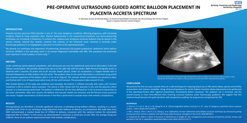
Under combined spinal-epidural anaesthetic, with ultrasound we scan the abdominal aorta and its bifurcation in the left oblique coronal plane. We partially displace the uterus to the right side with the probe. Right femoral retrograde access is obtained with a vascular US probe and an 8F vascular sheath placed. Under US visualisation a J-tipped metallic wire is inserted followed by an Atlas balloon into the aorta. The position close to the aortic bifurcation is confirmed using partial non-occlusive expansion of the balloon with 1−2 mL of air [Figure]. The vascular sheath and balloon are secured in place and flushed with 5 mL of heparinized saline every 10 min until removal. This procedure takes around 15 min.
After fundal delivery of the baby and umbilical cord clamping, the balloon is inflated manually with saline until minimal resistance is felt to achieve aortic occlusion. The uterus is then closed with the placenta in situ and the placenta either excised, or a hysterectomy performed. The balloon is inflated for 20 min then deflated for 5 min to preserve blood flow to the lower extremities. After skin closure, the balloon and vascular sheath are removed, and the femoral artery puncture site closed. Doppler examination of tibial arteries in the feet is performed to exclude lower limb embolic complications.
RESULTS
Intraoperatively we identified a clinically significant reduction in bleeding during balloon inflation, resulting in a clearer operating field. Prior to our technique using abdominal aortic balloons at delivery, our prospective PAS audit data since 2005 demonstrated a median blood loss of 2700ml at delivery, consistent with the national median for antenatally diagnosed PAS of 2750ml.3 In this series, we demonstrated a reduction in blood loss of over 50%, the average being just 1366mls. None of our patients experienced lower limb embolic complications.
CONCLUSION
We present the first six cases outside China of a safe technique for reducing blood loss in PAS which allows uterine and fertility preservation and reduces morbidity. Using ultrasound guidance for aortic balloon insertion allows placement to be performed in the operating theatre after regional anaesthesia, immediately before caesarean. The radiation risk is removed, and the overall process is more time-efficient than inserting occlusive balloons under fluoroscopic guidance. We suggest that this method should become the gold standard, with prospective auditing, for pregnancies complicated by PAS.
REFERENCES
1. Chen K, Li F, Jun E, Xie K, Li W, Zhang M, et al. Ultrasoundguided balloon occlusion in 13 cases of dangerous placental previa cesarean section. J Interv Radiol 2017;26:915-7.
2. Wei L, Gong G, Chen J, Hou P, Li Q, Zheng Z, et al. Application of lower abdominal aorta balloon occlusion technique by ultrasound guiding during caesarean section in patients with pernicious placenta previa. Zhonghua Yi Xue Za Zhi 2018;98:930-4.
3. Fitzpatrick KE, Sellers S, Spark P, Kurinczuk JJ, Brocklehurst P, Knight M. The management and outcomes of placenta accreta, increta, and percreta in the UK: A population-based descriptive study. BJOG 2014;121:62-71.