1,830 posters, 67 videos, 75 audios, 29 topics, 22 sessions, 4,727 authors, 1,697 institutions
ePostersLive by SciGen Technologies S.A. All rights reserved.
RCOG World Congress 2023
June 12 - 14, 2023 | London, UK

EP.1532
Routine Ultrasonography for Women of Childbearing Age? A Case of Uterine Didelphys
Poster Presenter
Authors
Affiliations
Part of Topic
Endometriosis and Uterine Disorders
Routine Ultrasonography for Women of Childbearing Age? A Case of Uterine Didelphys.
Introduction
Mullerian duct abnormalities (MDA) are a spectrum of congenital defects arising from failure of fusion of the Mullerian ducts during embryogenesis. They range between 0.5-5% of the general population. Uterine didelphys arises from the complete non-fusion of both Mullerian ducts and is one of the rarest forms of MDA’s. It is usually asymptomatic and therefore usually diagnosed during childbearing years as an incidental finding. During pregnancy it can be associated with increased rates of miscarriage, preterm delivery and growth restriction. Gold standard for diagnosis is the 3D ultrasonography with MRI being reserved for diagnosing complex MDA’s.
Case Report
History:
30-year-old female primigravida at 9+3 presented with a 5-day history of cramping abdominal pain and PV bleeding with clots and fleshy material.
Physical Exam:
Patiett had mild tenderness in the suprapubic region, minimal dark red blood in the vault and cervix opened to 2cm.
Investigations:
Urine beta HCG was positive, blood group B-positive and Hemoglobin was 13.2 mg/dL. Pelvic USS revealed No intrauterine pregnancy with mild retained products of conception in uterus.
Surgical Findings and Hospital Course
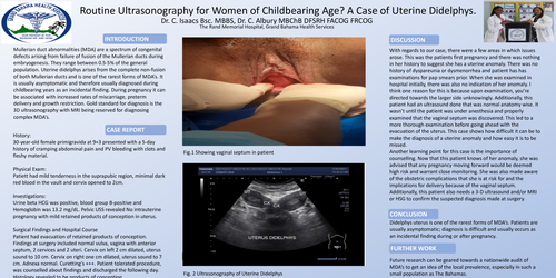
Patient had evacuation of retained products of conception. Findings at surgery included normal vulva, vagina with anterior septum, 2 cervices and 2 uteri. Cervix on left 2 cm dilated, uterus sound to 10 cm. Cervix on right one cm dilated, uterus sound to 7 cm. Adnexa normal. Curetting’s +++. Patient tolerated procedure, was counselled about findings and discharged the following day. Histology revealed to be products of conception.
Discussion
With regards to our case, there were a few areas in which issues arose. This was the patients first pregnancy and there was nothing in her history to suggest she has a uterine anomaly. There was no history of dyspareunia or dysmenorrhea and patient has has examinations for pap smears prior. When she was examined in hospital initially, there was also no indication of her anomaly. I think one reason for this is because upon examination, you’re directed towards the larger side unknowingly. Additionally, this patient had an ultrasound done that was normal anatomy wise. It wasn’t until the patient was under anesthesia and properly examined that the vaginal septum was discovered. This led to a more thorough examination before going ahead with the evacuation of the uterus. This case shows how difficult it can be to make the diagnosis of a uterine anomaly and how easy it is to be missed.
Another learning point for this case is the importance of counselling. Now that this patient knows of her anomaly, she was advised that any pregnancy moving forward would be deemed high risk and warrant close monitoring. She was also made aware of the obstetric complications that she is at risk for and the implications for delivery because of the vaginal septum. Additionally, this patient also needs a 3-D ultrasound and/or MRI or HSG to confirm the suspected diagnosis made at surgery.
Conclusion
Didelphys uterus is one of the rarest forms of MDA’s. Patients are usually asymptomatic; diagnosis is difficult and usually occurs as an incidental finding during or after pregnancy.
Further Work
Future research can be geared towards a nationwide audit of MDA’s to get an idea of the local prevalence, especially in such a small population as The Bahamas.