1,830 posters, 67 videos, 75 audios, 29 topics, 22 sessions, 4,727 authors, 1,697 institutions
ePostersLive by SciGen Technologies S.A. All rights reserved.
RCOG World Congress 2023
June 12 - 14, 2023 | London, UK

EP.1367
Pregnancy in Uterus bicornis unicollis misdiagnosed as an ectopic pregnancy.
Poster Presenter
Part of Topic
Endometriosis and Uterine Disorders
Pregnancy in Uterus bicornis unicollis misdiagnosed as ectopic pregnancy
Dr Bilques Bilques, Dr Judith Docherty, Mr Rahul Savant, Mr Samendra Roy, Dr Mihraban Bapir
North Tees and Hartlepool NHS Foundation trust
Case report:
A 32 years old lady G2 P0 at 6 weeks of pregnancy presented with vaginal bleeding. She had previous medical termination. She suffered from anxiety but was otherwise fit and well with no previous surgery. Scan in early pregnancy clinic showed a live ectopic pregnancy of 6 weeks gestation in the right adnexa with a normal anteverted uterus. we consented her for diagnostic Laparoscopy and management of ectopic pregnancy.
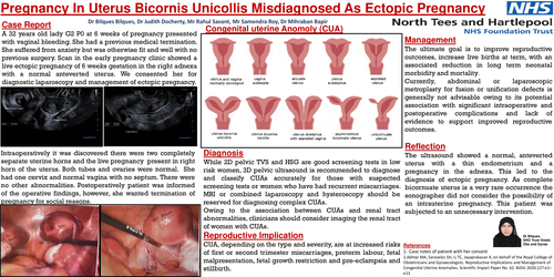
Intraoperatively it was discovered that she has two completely separate uterine horns and the live pregnancy present in right horn of the uterus. Her both tubes and ovaries were normal. She had one cervix and normal vagina with no septum. There were no other abnormalities. Postoperatively she was informed of the operative findings, however she wanted termination of pregnancy for social reasons.
Discussion:
Anatomy
The female genital tract arises from two embryological elements, the Mullerian ducts and the urogenital sinus. The Mullerian ducts differentiate to form the fallopian tubes, body of the uterus, cervix and upper part of the vagina. The urogenital sinus gives rise to the mid and lower part of the vagina. Uterus bicornis unicollis is due to failure of midline fusion of the Mullerian ducts to form a single uterus body and cervix. Pregnancy in an anomalous uterus is high risk.
Diagnosis
While 2D pelvic TVS and HSG are good screening tests in low risk women, 3D pelvic ultrasound is recommended to diagnose and classify CUAs accurately for those with suspected screening tests or women who have had recurrent miscarriages. MRI or combined laparoscopy and hysteroscopy should be reserved for diagnosing complex CUAs.
Owing to the association between CUAs and renal tract abnormalities, clinicians should consider imaging the renal tract of women with CUAs.
Reproductive Implication
CUA, depending on the type and severity, are at increased risks of first or second trimester miscarriages, preterm labour, fetal malpresentation, fetal growth restriction and pre-eclampsia and stillbirth.
Management
The ultimate goal is to improve reproductive outcomes, increase live births at term, with an associated reduction in long term neonatal morbidity and mortality.
Currently, abdominal or laparoscopic metroplasty for fusion or unification defects is generally not advisable owing to its potential association with significant intraoperative and postoperative complications and lack of evidence to support improved reproductive outcomes.
Reflection:
The ultrasound showed a normal, anteverted uterus with a thin endometrium and a pregnancy in the adnexa. This led to the diagnosis of ectopic pregnancy. As complete bicornuate uterus is a very rare occurrence the sonographer did not consider the possibility of an intrauterine pregnancy. This patient was subjected to an unnecessary intervention.