1,830 posters, 67 videos, 75 audios, 29 topics, 22 sessions, 4,727 authors, 1,697 institutions
ePostersLive by SciGen Technologies S.A. All rights reserved.
RCOG World Congress 2023
June 12 - 14, 2023 | London, UK

EP.1244
A case with bicornuate uterus and solitary kidney with complete duplex system
Poster Presenter
Authors
Affiliations
Part of Topic
Endometriosis and Uterine Disorders
Video
Title:
A case with bicornuate uterus and solitary kidney with complete duplex system.
Objective:
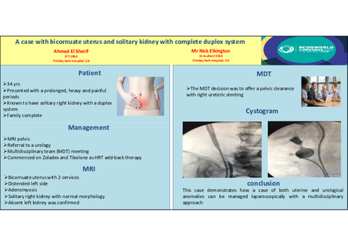
To share an interesting case, by presenting a video showing a total laparoscopic hysterectomy with right salpingectomy and bilateral oophorectomy, for a patient diagnosed with a bicornuate uterus and single right kidney with double ureters.
Design:
In this video presentation we show an edited video first showing cystoscopy with stenting of the 2 ureteric orifices, and retrograde pictures demonstrating duplex ureters draining the right kidney. This was followed by laparoscopic adhesiolysis to access the right adnexa, and then a total laparoscopic hysterectomy with right salpingectomy, bilateral oophorectomy and removal of two cervices.
Method:
A 34-year-old woman presented with a prolonged, heavy and painful periods. At the age of 15 she had a left laparoscopic salpingectomy for a torted left fallopian tube. She was known to have a congenitally absent left kidney. She had previously had one social termination of pregnancy and had firmly decided that she never wanted to have children. She was initially thought to have endometriosis and was started on Zoladex and Tibolone as HRT add-back therapy.
Results:
Examination and transvaginal ultrasound (US) scan showed a fixed tender bicornuate uterus, one cervix, and normal ovaries. Further evaluation by US scan and MRI confirmed a bicornuate uterus with 2 cervices with a distended left side and adenomyosis was suspected. Also, a solitary right kidney with normal morphology and an absent left kidney was confirmed. Her ovaries, liver, gall bladder, pancreas and spleen were all normal. Following an endometriosis MDT, it was decided to perform a pelvic clearance with right ureteric stenting. A pre-operative urology review recommended a CT urogram, which confirmed a solitary right kidney with a duplex system.
Conclusions
This case demonstrates how a case of both uterine and urological anomalies can be managed laparoscopically with a multidisciplinary approach.