215 posters, 5 topics, 22 sessions, 596 authors, 283 institutions
ePostersLive by SciGen Technologies S.A. All rights reserved.
Obstetric Anaesthesia Annual Scientific Meeting 2025
May 15 - 16, 2025 | ICC, 2 Lanyon Place, Belfast BT1 3WH

6779704
Measured blood loss - the casualty of cell salvage
Poster Presenter
Part of Topic
Service Evaluation
Introduction
It has become common practice to employ gravimetric recording of blood loss during Caesarean sections—i.e., calculating the total wet weight of swabs plus the suction reservoir, minus the dry weight of the swabs. The accuracy of this method, its comparison with photometric techniques, and its validation against Brecher’s formula [1] have been the subject of numerous studies and reviews [2].
The use of autotransfusion (“cell salvage”) introduces additional variables into blood loss calculation. At our institution, blood and amniotic fluid are collected directly into the cell salvage reservoir, mixing with anticoagulant. The surgical swabs are washed in the sterile field as soon as practical and collected in batches into the reservoir - limiting real time totals of blood loss. We process using a Fresenius Kabi CATSmart® machine.
Swabbed blood is exposed to pressure and foreign surfaces without anticoagulation, likely leading to clotting and haemolysis. Cell salvage efficiency is never 100%, but the impact of this additional error is unknown. Ideally, reservoir and swab blood would be processed separately to test this, but delaying processing until the end of surgery is impractical.
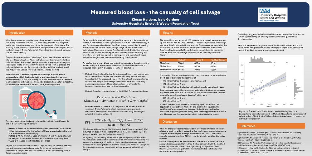
There are two main methods currently used to estimate blood loss at the end of a case involving cell salvage:
As part of a service audit of our cell salvage practice, we aimed to compare how well these two methods correlate. To do so, we performed a retrospective analysis of blood loss estimates over a four-month period of Caesarean section cases.
Methods
We surveyed the hospitals in our geographical region and determined that Method 1 and Method 2 were equally utilized, with no third methodology in use. We retrospectively collected data from January to April 2024, drawing from hand-written records of cell salvage usage, as well as electronic maternity and laboratory systems. The dataset included collection volumes, amniotic fluid volume, swab weights, fluid volumes introduced during the procedure, pre- and post-operative haemoglobin and haematocrit values, and patient weight (used to estimate circulating blood volume).
We applied two primary blood loss estimation methods to the retrospective dataset, along with a comparator calculation (Modifed Brecher) based on measured haemoglobin changes pre- and post-transfusion:
- Method 1 involved multiplying the autologous blood return volume by a factor derived from the machine's quoted efficiency and the average haematocrit of the processed output [3]. This calculation was performed twice—once using a fixed average haematocrit value and once using patient-specific measured haematocrit values—to eliminate the haematocrit percentage as a confounding variable.
- Method 2 used an equation based on the UK Cell Salvage Action Group
[Equation 1]
[Equation 2]
Recognising that assuming a haematocrit percentage may introduce error, we conducted a spot audit in December 2024. This involved measuring the haematocrit percentage of 10 blood return samples processed using all machines in use during that year. We then recalculated Method 1 using both the manufacturer-quoted haematocrit and our experimentally derived average.
Results
The mean blood loss across all 265 patients for whom cell salvage was set up was 546 ml (SD: 497 ml). Of these, 77 cases had processed cell salvage and were therefore included in our analysis. Eleven cases were excluded due to concomitant donor blood transfusions (which rendered the modified Brecher calculation unreliable), and six were excluded due to incomplete data. As expected, the average blood loss in the remaining cases was higher.
[Table 1]
The modified Brecher equation indicated that both methods underestimated blood loss, with average discrepancies of:
Since these are mean differences, over- and underestimations across cases may cancel each other out. To account for this, we also calculated absolute mean differences (regardless of direction), which were:
A paired samples t-test showed a statistically significant difference in population means between Method 1 and the Brecher equation. No significant difference was found between Method 2 and the Brecher calculation, suggesting Method 2 may more closely approximate true blood loss. However, this finding may also reflect limited statistical power.
[Figure 1]
Discussion
While we anticipated some inaccuracies in blood loss estimation when cell salvage is used, we did not expect the degree of error observed with widely accepted methodologies. Average discrepancies of 122–173 ml—and absolute errors approaching 400 ml—may be clinically significant in certain contexts.
Despite its complexity, we will continue to use Method 2. In our dataset, it appeared more accurate than Method 1 when compared with the modified Brecher equation and did not differ significantly in population mean. However, we acknowledge that this may reflect limited statistical power rather than a true equivalence.
Our findings suggest that both methods introduce measurable error, and we caution against relying on any single absolute value to guide clinical decision-making.
Method 2 has potential to give an earlier final loss calculation, as it is not reliant on the final processed volume. Attempts to improve the accuracy of Method 2 are likely to come at the expense of speed.