708 posters, 8 topics, 16 sessions, 1,190 authors, 600 institutions
ePostersLive by SciGen Technologies S.A. All rights reserved.
ISQua's 39th International Conference 2023
August 27 - 30, 2023 | Seoul, Republic of Korea

P014
COMPARISON OF DETECTION OF SUPERIOR GLUTEAL ARTERY PERFORATOR BY INDOCYANINE GREEN FLUORESCENCE NEAR-INFRARED ANGIOGRAPHY AND ACOUSTIC DOPPLER SONOGRAPHY FOR RECONSTRUCTION OF SACRAL PRESSURE INJURY
Part of Topic
Digital Healthcare and Innovation
COMPARISON OF DETECTION OF SUPERIOR GLUTEAL ARTERY PERFORATOR BY INDOCYANINE GREEN
FLUORESCENCE NEAR-INFRARED ANGIOGRAPHY AND ACOUSTIC DOPPLER SONOGRAPHY FOR RECONSTRUCTION OF SACRAL PRESSURE INJURY
Chien-Wei Wu and Yuan-Sheng Tzeng
Division of Plastic and Reconstruction Surgery, Department of Surgery, Tri-Service General Hospital, National Defense Medical Center, Taipei, Taiwan
Introduction
Pressure injuries are areas with tissue necrosis and ulceration where soft tissues are compressed between hard surfaces and bony structure. Once deep tissue infection is noted, in-hospital care, along with surgical interventions, is beneficial for patients. Several different kinds of flaps have been introduced over the years for reconstructing sacral defects, such as local flaps, V-Y advancement fasciocutaneous flaps, gluteus maximum muscle-based flaps, inferior gluteal artery perforator flaps, and superior gluteal artery perforator (SGAP) flaps.Reliable perforators are the most important factors for successful perforator based flaps reconstruction
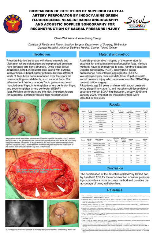
A hypothetical line was drawn between the posterior superior iliac spine (PSIS) and the lateral border of the great trochanter on the side of the buttock from which the SGAP flap was planned to be harvested. B) A hypothetical line was drawn between the posterior superior iliac spine (PSIS) and the lateral border of the great trochanter on the side of the buttock from which the SGAP flap was be harvested.
The superior gluteal artery (SGA) and its perforators were identified with ICGFA and ADS.
Material and method
Accurate preoperative mapping of the perforators is essential for the safe planning of propeller flaps. Various methods have been reported to date: handheld acoustic Doppler sonography (ADS), indocyanine green fluorescence near-infrared angiography (ICGFA)
We retrospectively reviewed data from 16 patients with sacral pressure injury who underwent modified SGAP flap reconstruction surgery.
All patients age 65 years and over with sacral pressure injury stage III to stage IV, and massive soft tissue defect coverage with an SGAP flap between January 2019 and January 2021, who met the inclusion criteria were
included in this study.
Results
SGAP flap was tunnelled beneath a skin strip between the defect and the flap donor site.
Conclusion
The combination of the detection of SGAP by ICGFA and by handheld ADS for the reconstruction of sacral pressure injury provides a more accurate method and provides the advantage of being radiation-free.
Reference
-
Khurram, M.F.; Khan, A.H.; Ahmad, I.; Nanda, M.; Masoodi, Z. Superior gluteal artery perforator flap: A reliable method for sacral pressure ulcer reconstruction. J. Wound Care 2013, 22, 699–703.
-
Chang, C.K.; Wu, C.J.; Chen, C.Y.; Wang, C.Y.; Chu, T.S.; Hsu, K.F.; Chiu, H.T.; Liu, H.H.; Chou, C.Y.; Wang, C.H.; et al. Intraopera- tive indocyanine green fluorescent angiography-assisted modified superior gluteal artery perforator flap for reconstruction of sacral pressure sores. Int. Wound J. 2017, 14, 1170–1174.
-
Onoda, S.; Azumi, S.; Hasegawa, K.; Kimata, Y. Preoperative identification of perforator vessels by combining MDCT, doppler flowmetry, and ICG fluorescent angiography. Microsurgery 2013, 33, 265–269.
-
Ritschl, L.M.; Schmidt, L.H.; Fichter, A.M.; Hapfelmeier, A.; Wolff, K.D.; Mucke, T. Multimodal analysis using flowmeter analysis, laser- Doppler spectrophotometry, and indocyanine green videoangiography for the detection of venous compromise in flaps in rats. J. Cranio- Maxillofac. Surg. 2018, 46, 905–915.
-
Lin, C.T.; Chang, S.C.; Chen, S.G.; Tzeng, Y.S. Modification of the superior gluteal artery perforator flap for reconstruction of sacral sores. J. Plast. Reconstr. Aesthet. Surg. 2014, 67, 526–532.
-
Georgiou, A.K.; Singh, P.; Hever, P.; Arize, C.; Mosahebi, A. The use of indocyanine green in plastic surgery. J. Plast. Reconstr. Aesthet. Surg. 2020, 73, e8–e9
-
Holm, C.; Mayr, M.; Hofter, E.; Becker, A.; Pfeiffer, U.J.; Muhlbauer, W. Intraoperative evaluation of skin-flap viability using laser-induced fluorescence of indocyanine green. Br. J. Plast. Surg. 2002, 55, 635–644.
-
Ritschl, L.M.; Fichter, A.M.; Bomhard, A.V.; Koerdt, S.; Kehl, V.; Kolk, A.; Wolff, K.D.; Grill, F.D. Comparison between Different Perforator Imaging Modalities for the Anterolateral Thigh Perforator Flap Transfer: A Prospective Study. J. Reconstr. Microsurg. 2020, 36, 686–693.
-
Mucke, T.; Hapfelmeier, A.; Schmidt, L.H.; Fichter, A.M.; Kanatas, A.; Wolff, K.D.; Ritschl, L.M. A comparative analysis using flowmeter, laser-Doppler |spectrophotometry, and indocyanine green-videoangiography for detection of vascular stenosis in free flaps. Sci. Rep. 2020, 10, 939