301 posters, 50 videos, 13 topics, 13 sessions, 734 authors, 193 institutions
ePostersLive by SciGen Technologies S.A. All rights reserved.
ISMICS 2026 Annual Meeting
10 - 13 June, 2026 | Miami, Florida

D78
Feasibility Study Of Histological Damage In Endoscopic Long Saphenous Vein Harvesting During The Learning Curve
Poster Presenter
Authors
Sam Raaj, Rick Air, Cristina Ruiz Segria, Setemi Olufemi, Azita Rajai, Bhuvaneswari Krishnamoorthy
Affiliations
Directorate of Nursing, Midwifery & Social Care, University of Salford, England, United Kingdom, Faculty of Medicine, Medical School, Imperial College of London, England, United Kingdom, Department of Cardiothoraic Surgery, Manchester Foundation Trust, Manchester, England, United Kingdom, Department of Cardiothoracic Surgery, St. James Cook hospital, SouthTeeside, England, United Kingdom, Department of Medical Statistics, University of Manchester, England, United Kingdom
Part of Topic
Coronary Revascularization
Background
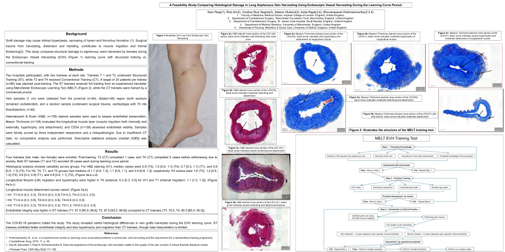
Graft damage may cause intimal hyperplasia, narrowing of lumen and thrombus formation (1). Surgical trauma from harvesting, distension and handling, contributes to muscle migration and intimal thickening(2). This study compares structural damage to saphenous veins harvested by trainees during the Endoscopic Vessel Harvesting (EVH) (Figure 1) learning curve with structured training vs. conventional training.
Methods
Two hospitals participated, with two trainees at each site. Trainees T 1 and T2 underwent Structured Training (ST), while T3 and T4 received Conventional Training (CT). A target of 20 patients per trainee (n=80) was planned post-training. The ST trainees received full training from an experienced harvester using Manchester Endoscopic Learning Tool (MELT) (Figure 2), while the CT trainees were trained by a commercial proctor.
Vein samples (1 cm) were collected from the proximal (n=46), distal(n=46) region (both sections remained undistended), and a random sample (underwent surgical trauma, cardioplegia with 70 mls flow/distention, n=46).
Haematoxylin & Eosin (H&E, n=138) stained samples were used to assess endothelial preservation, Mason Trichrome (n=138) evaluated the longitudinal muscle layer (muscle migration both internally and externally, hypertrophy, and detachment), and CD34 (n=138) assessed endothelial viability. Samples were blindly scored by three independent researchers and a histopathologist. Due to insufficient CT data, no comparative analysis was performed. Descriptive statistical analysis (median [IQR]) was calculated.
Results
Four trainees (two male, two female) were enrolled. Post-training, T3 (CT) completed 1 case, and T4 (CT) completed 5 cases before withdrawing due to anxiety. Both ST trainees (T1 and T2) recruited 20 cases each during learning curve period.
Histological analysis showed variability across groups. For H&E staining (H1), median values were 0.9 (T3), 1.0 [0.9, 1.1] (T4), 0.7 [0.3, 1.1] (T1), and 0.8 [0.6, 1.1] (T2). For H2, T4, T1, and T2 groups had medians of 1.1 [0.9, 1.2], 1.1 [0.9, 1.1], and 0.9 [0.8, 1.2], respectively. H3 scores were 1.6 (T3), 1.2 [0.9, 1.2] (T4), 0.6 [0.3, 0.9] (T1), and 0.8 [0.6, 1.1] (T2). (Figure 3a,b,c,d)
Longitudinal Muscle (LM) migration and hypertrophy were higher in T4 (external: 0.3 [0.3, 0.5] for H1) and T1 (internal migration: 1.1 [1.0, 1.3]). (Figure 4a,b,c)
Longitudinal muscle detachment scores varied: (Figure 5a,b)
• H1: T1=0.0 (0.0, 0.3), T2=0.0 (0.0, 0.5) T3=0.3, T4=0.3 (0.3, 0.5)
• H2: T1=0.4 (0.3, 0.8), T2=0.0 (0.0, 0.8), T4=0.6 (0.3, 0.9)
• H3: T1=0.0 (0.0, 0.3), T2=0.0 (0.0, 0.3), T3=1.3, T4=0.5 (0.3, 0.5)
Endothelial integrity was higher in ST trainees (T1: 97.5 [95.9, 98.6], T2: 97.5 [93.5, 98.8]) compared to CT trainees (T3: 75.0, T4: 85.0 [85.0, 90.5]).
Conclusion
The COVID-19 pandemic halted the study. The study revealed varied histological differences in vein grafts harvested during the EVH learning curve. ST trainees exhibited better endothelial integrity and less hypertrophy and migration than CT trainees, though data interpretation is limited.