301 posters, 50 videos, 13 topics, 13 sessions, 734 authors, 193 institutions
ePostersLive by SciGen Technologies S.A. All rights reserved.
ISMICS 2026 Annual Meeting
10 - 13 June, 2026 | Miami, Florida

D15
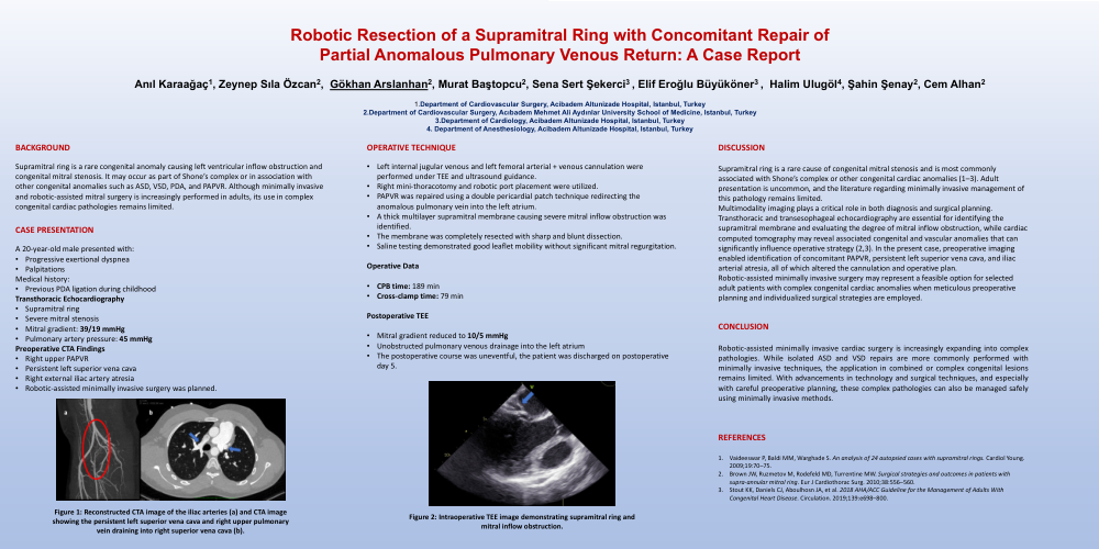
Robotic Resection Of A Supramitral Ring With Concomitant Repair Of Partial Anomalous Pulmonary Venous Return: A Case Report
Poster Presenter
Authors
Anıl Karaağaç, Zeynep Sıla Özcan, Gökhan Arslanhan, Murat Baştopcu, Sena Sert Şekerci, Elif Eroğlu Büyüköner, Halim Ulugöl, Şahin Şenay, Cem Alhan
Affiliations
Department of Cardiovascular Surgery, Acibadem Altunizade Hospital, Istanbul, Turkey, Department of Cardiovascular Surgery, Acıbadem Mehmet Ali Aydınlar University School of Medicine, Istanbul, Turkey, Department of Cardiology, Acibadem Altunizade Hospital, Istanbul, Turkey, Department of Anesthesiology, Acibadem Altunizade Hospital, Istanbul, Turkey
Part of Topic
Valve � Surgical Valves
Video
BACKGROUND
Supramitral ring is a rare congenital anomaly causing left ventricular inflow obstruction and congenital mitral stenosis. It may occur as part of Shone’s complex or in association with other congenital anomalies such as ASD, VSD, PDA, and PAPVR. Although minimally invasive and robotic-assisted mitral surgery is increasingly performed in adults, its use in complex congenital cardiac pathologies remains limited.
CASE PRESENTATION
A 20-year-old male presented with:
• Progressive exertional dyspnea
• Palpitations
Medical history:
• Previous PDA ligation during childhood
Transthoracic Echocardiography
-
Supramitral ring
-
Severe mitral stenosis
-
Mitral gradient: 39/19 mmHg
-
Pulmonary artery pressure: 45 mmHg
Preoperative CTA Findings
-
Right upper PAPVR
-
Persistent left superior vena cava
-
Right external iliac artery atresia
-
Robotic-assisted minimally invasive surgery was planned
OPERATIVE TECHNIQUE
• Left internal jugular venous and left femoral arterial + venous cannulation were performed under TEE and ultrasound guidance.
• Right mini-thoracotomy and robotic port placement were utilized.
• PAPVR was repaired using a double pericardial patch technique redirecting the anomalous pulmonary vein into the left atrium.
• A thick multilayer supramitral membrane causing severe mitral inflow obstruction was identified.
• The membrane was completely resected with sharp and blunt dissection.
• Saline testing demonstrated good leaflet mobility without significant mitral regurgitation.
Operative Data
• CPB time: 189 min
• Cross-clamp time: 79 min Postoperative TEE
-
Mitral gradient reduced to 10/5 mmHg
-
Unobstructed pulmonary venous drainage into the left atrium
-
The postoperative course was uneventful, the patient was discharged on postoperative day 5.
DISCUSSION
Supramitral ring is a rare cause of congenital mitral stenosis and is most commonly associated with Shone’s complex or other congenital cardiac anomalies (1–3). Adult presentation is uncommon, and the literature regarding minimally invasive management of this pathology remains limited.
Multimodality imaging plays a critical role in both diagnosis and surgical planning. Transthoracic and transesophageal echocardiography are essential for identifying the supramitral membrane and evaluating the degree of mitral inflow obstruction, while cardiac computed tomography may reveal associated congenital and vascular anomalies that can significantly influence operative strategy (2,3). In the present case, preoperative imaging enabled identification of concomitant PAPVR, persistent left superior vena cava, and iliac arterial atresia, all of which altered the cannulation and operative plan.
Robotic-assisted minimally invasive surgery may represent a feasible option for selected adult patients with complex congenital cardiac anomalies when meticulous preoperative planning and individualized surgical strategies are employed.
CONCLUSION
Robotic-assisted minimally invasive cardiac surgery is increasingly expanding into complex pathologies. While isolated ASD and VSD repairs are more commonly performed with minimally invasive techniques, the application in combined or complex congenital lesions remains limited. With advancements in technology and surgical techniques, and especially with careful preoperative planning, these complex pathologies can also be managed safely using minimally invasive methods.
REFERENCES
-
Vaideeswar P, Baldi MM, Warghade S. An analysis of 24 autopsied cases with supramitral rings. Cardiol Young. 2009;19:70–75.
-
Brown JW, Ruzmetov M, Rodefeld MD, Turrentine MW. Surgical strategies and outcomes in patients with supra-annular mitral ring. Eur J Cardiothorac Surg. 2010;38:556–560.
-
Stout KK, Daniels CJ, Aboulhosn JA, et al. 2018 AHA/ACC Guideline for the Management of Adults With Congenital Heart Disease. Circulation. 2019;139:e698–800.