293 posters, 56 videos, 24 topics, 13 sessions, 658 authors, 215 institutions
ePostersLive by SciGen Technologies S.A. All rights reserved.
ISMICS 2025 Annual Meeting
28 - 31 May, 2025 | Istanbul, Turkey

D109
Robotically-assisted Aortic Valve Replacement: A Case Report
Poster Presenter
Part of Topic
Valve – Surgical Valves
Video
Introduction
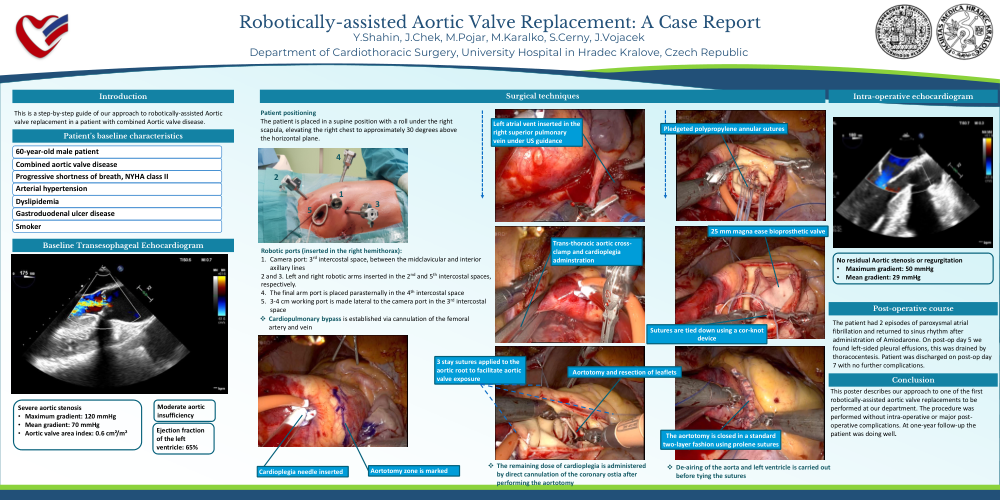
This is a step-by-step guide of our approach to robotically-assisted Aortic
valve replacement in a patient with combined Aortic valve disease.
Patient’s baseline characteristics
60-year-old male patient
Combined aortic valve disease
Progressive shortness of breath, NYHA class II
Arterial hypertension
Dyslipidemia
Gastroduodenal ulcer disease
Smoker
Baseline Transesophageal Echocardiogram
Severe aortic stenosis
• Maximum gradient: 120 mmHg
• Mean gradient: 70 mmHg
• Aortic valve area index: 0.6 cm²/m²
Moderate aortic insufficiency
Ejection fractionof the left ventricle: 65%
Surgical techniques
Patient positioning
The patient is placed in a supine position with a roll under the right
scapula, elevating the right chest to approximately 30 degrees above
the horizontal plane.
Robotic ports (inserted in the right hemithorax):
1. Camera port: 3rd intercostal space, between the midclavicular and interior
axillary lines
2 and 3. Left and right robotic arms inserted in the 2nd and 5th intercostal spaces,
respectively.
4. The final arm port is placed parasternally in the 4th intercostal space
5. 3-4 cm working port is made lateral to the camera port in the 3rd intercostal
space
❖ Cardiopulmonary bypass is established via cannulation of the femoral
artery and vein
Cardioplegia needle inserted
Aortotomy zone is marked
Left atrial vent inserted in the right superior pulmonary vein under US guidance
Trans-thoracic aortic crossclamp and cardioplegia adminstration
Aortotomy and resection of leaflets
3 stay sutures applied to the aortic root to facilitate aortic
valve exposure
❖ The remaining dose of cardioplegia is administered by direct cannulation of the coronary ostia after performing the aortotomy
Pledgeted polypropylene annular sutures
25 mm magna ease bioprosthetic valve
Sutures are tied down using a cor-knot device
The aortotomy is closed in a standard two-layer fashion using prolene sutures
❖ De-airing of the aorta and left ventricle is carried out before tying the sutures
Intra-operative echocardiogram
No residual Aortic stenosis or regurgitation
• Maximum gradient: 50 mmHg
• Mean gradient: 29 mmHg
Post-operative course
The patient had 2 episodes of paroxysmal atrial fibrillation and returned to sinus rhythm after administration of Amiodarone. On post-op day 5 we found left sided pleural effusions, this was drained by thoracocentesis. Patient was discharged on post-op day 7 with no further complications.
Conclusion
This poster describes our approach to one of the first robotically-assisted aortic valve replacements to be performed at our department. The procedure was
performed without intra-operative or major postoperative complications. At one-year follow-up the patient was doing well.